
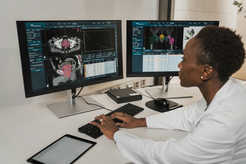
Imagine trying to understand how a watch works while blindfolded, allowed only to shake it and listen to the ticking. That was medicine, engineering, and much of science for most of human history. We could poke, prod, and guess, but actually seeing the inside of living bodies or complex machines in real time was almost impossible without breaking them open.
Then, slowly at first and now at breakneck speed, something radical happened: we learned how to . X‑rays, ultrasound, CT scans, MRI, endoscopy, PET, functional imaging, high‑resolution microscopy, non‑invasive sensors – together they flipped the scientific world inside out. We stopped just treating symptoms and began seeing processes. We stopped guessing and started watching, live.
This shift did not just make things safer. It changed what questions researchers even thought to ask, how they tested their ideas, and how quickly they could be proven wrong. It rewired how we train doctors, how we design drugs, how we study the brain, and even how we think about privacy and what it means to be “healthy.”
Let’s walk through what really changed once we could peer under the skin and into the hidden workings of bodies, brains, and machines – without a single incision.
#1 From Guessing To Seeing: How Non‑Invasive Imaging Rewired Diagnosis
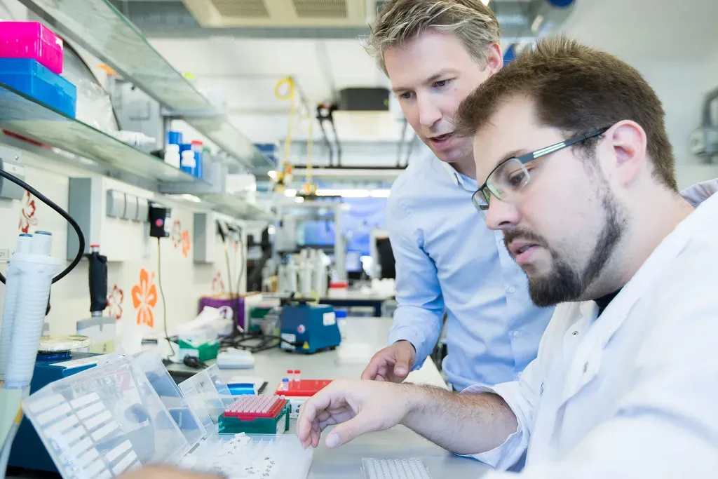
It’s hard to overstate how much of early medicine was educated guesswork. Doctors listened, tapped, and felt, then tried to imagine what might be wrong inside. Autopsies sometimes confirmed their ideas, but only after it was too late to help. The first X‑ray images at the end of the nineteenth century were a shock: suddenly you could see bones, bullets, and large lung lesions in living people, almost like having a crude superpower.
Over the twentieth century that crude superpower became a toolbox. X‑rays were joined by ultrasound, CT, MRI, angiography, and nuclear scans. Diagnosis moved from “you probably have…” to “here is the exact size, shape, and position of the problem.” Instead of inferring a fracture from pain and deformity, you could see the broken bone. Instead of guessing whether a stroke was bleeding or a clot, you could see the brain’s damaged region and its blood vessels in detail.
This shift didn’t just improve accuracy; it changed the entire rhythm of care. Diseases that once required exploratory surgery to confirm, like certain tumors or abdominal catastrophes, could be seen non‑invasively. That meant faster treatment, fewer dangerous procedures, and a different psychological experience for patients – more evidence, less mystery, and often less fear that everything was a coin toss.
Today, a modern hospital would be almost unrecognizable without its imaging suites. For many conditions, the question is no longer “should we image?” but “which kind of imaging gives us the clearest answer with the least risk?” It’s a subtle but massive philosophical turn: when you can look, you stop telling stories and start showing pictures.
#2 Safer Science: When Cutting Became Plan B Instead Of Plan A
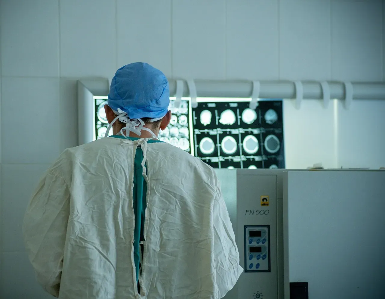
Before researchers could look inside safely, understanding organs and diseases often meant cutting first and asking questions later. Exploratory surgery – literally opening someone up to see what was wrong – was once common. That approach was brave, but also brutal: high risk, painful recoveries, and sometimes little to show for it beyond “we didn’t find much.”
Non‑invasive imaging flipped that order. Cutting became plan B. Now, surgeons routinely map out operations in advance using CT or MRI scans, tracing critical blood vessels, nerves, and tumor margins on a screen before making the first incision. That kind of planning shrinks operating times, reduces complications, and lets teams practice difficult cases virtually.
From a research perspective, this also opened up ethical territory that used to be off‑limits. You can scan a person repeatedly over time to watch how a disease develops, how an organ recovers, or how a new treatment affects the body. Longitudinal studies that would have been unthinkable if they required repeated surgical sampling became feasible and acceptable.
In practice, this has meant fewer emergency “open and see” surgeries, more minimally invasive procedures guided by images, and a culture where the scalpel is no longer the primary diagnostic tool. The knife did not disappear – but it became far more informed, precise, and often secondary to what the images first revealed.
#3 Precision In Real Time: Guiding Treatment From The Inside Out
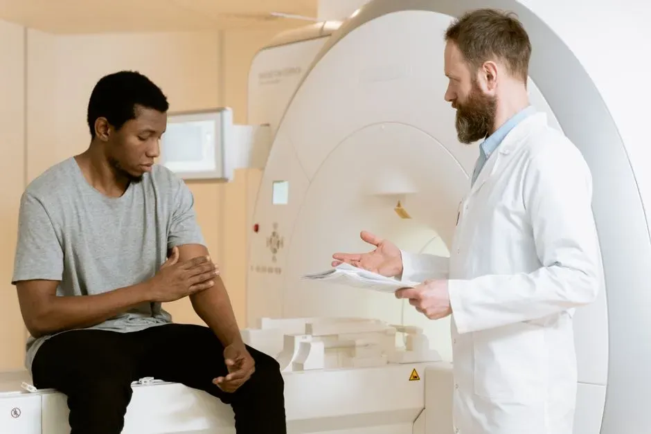
Once you can see inside without cutting, the obvious next step is to treat while you are watching. That idea gave rise to image‑guided interventions: using imaging in real time to steer needles, catheters, and tiny instruments exactly where they need to go, turning what used to be big surgeries into small punctures and brief procedures.
Interventional radiology is the clearest expression of this shift. Clot‑busting devices are threaded through arteries to open blocked vessels in the brain during a stroke, guided by live X‑ray images. Small coils or plugs are placed inside bulging blood vessels to prevent deadly ruptures, with every move tracked on the screen. Tumors in the liver can be attacked from within by delivering chemotherapy or heat directly into the lesion while CT or ultrasound confirms that the target is hit.
Other specialties followed the same logic. Cardiologists use imaging to place stents, replace heart valves, and assess blood flow without opening the chest. Pain specialists place nerve blocks under ultrasound guidance rather than guessing based on anatomy diagrams. Even radiation therapy for cancer now uses detailed CT and MRI planning to shape beams that conform around tumors and spare healthy tissues as much as possible.
In daily practice, this means treatments that are often:
- More precise, because clinicians see exactly where tools and drugs are going.
- Less invasive, relying on tiny entry points instead of large incisions.
- Faster to recover from, with many procedures done in hours instead of days.
The technology did not just help us understand what was wrong – it became the eyes and hands of a new kind of therapy.
#4 Seeing The Invisible: Early Disease, Subtle Changes, And The Age Of Screening

One of the most profound changes after we learned to look inside without cutting was simply this: we started catching things long before they caused trouble. Many diseases do their worst damage quietly. Tumors grow in silence. Arteries slowly narrow. Bone thins. Brains lose cells long before memory obviously slips. Without imaging and other non‑invasive tools, most of that remained invisible until it was almost too late.
Screening programs grew out of this new visibility. Mammography seeks breast cancers when they are still small and localized. Low‑dose CT in high‑risk smokers aims to find lung cancers while surgery is still an option. Bone density scans flag osteoporosis years before the first fracture. Cardiac imaging and calcium scoring spot plaque in coronary arteries before a heart attack announces itself with chest pain.
Of course, this early vision brought nuance and controversy. Seeing more does not automatically mean helping more. Some slow‑growing cancers might never have caused harm in a person’s lifetime, yet once discovered they can trigger anxiety, biopsies, and treatments. Researchers have had to wrestle with overdiagnosis – finding abnormalities that look scary but do not behave aggressively – and refine guidelines about when the benefits of early detection outweigh the harms.
Still, the net effect has been a head start. Instead of waiting for catastrophic symptoms, medicine can now often intervene earlier, when options are broader and outcomes better. On an emotional level, it has also transformed how many people think about their own bodies: not just as ticking time bombs, but as systems that can be monitored, scanned, and nudged away from disaster before it strikes.
#5 A New Window Into The Brain: From Black Box To Living Network
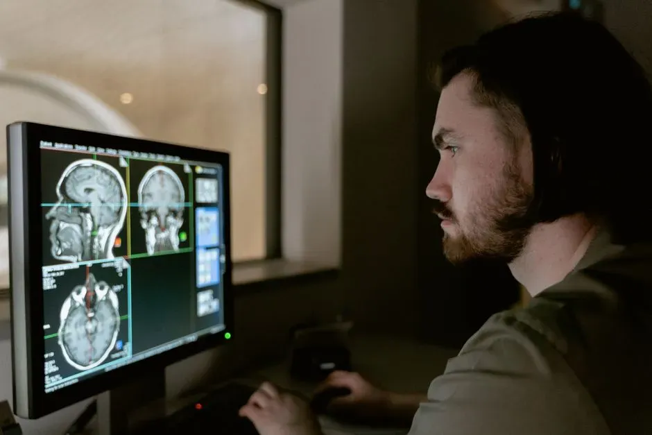
For centuries, the human brain was the ultimate black box. You could study injured soldiers, do autopsies, or perform crude electrical stimulation during open‑brain surgery, but there was no way to watch a healthy brain think, feel, or remember in real time. Non‑invasive brain imaging changed that, and with it, our entire sense of what neuroscience could be.
Computed tomography and later MRI first offered detailed structural views of the brain: tumors, strokes, malformations, and shrinking areas in degenerative diseases. That alone transformed neurology, letting clinicians see exactly where and how brain tissue was damaged. But the real revolution came when techniques like functional MRI and PET allowed researchers to measure blood flow and metabolism as proxies for neural activity in awake, behaving people.
Suddenly, it became possible to ask questions that once would have sounded like science fiction. What patterns of brain activity accompany language, decision‑making, or emotional reactions? How does addiction alter reward circuits? Which areas change as children learn to read, or as older adults start to slip into dementia? Large research programs sprang up around these questions, mapping networks instead of just isolated “centers.”
Of course, brain imaging has limitations. The colorful activation maps you see in popular media can be oversold, and brain activity is vastly more complex than a few highlighted blobs on a scan. But the core change is real and irreversible: the brain is no longer an unreachable mystery organ. It is a living, measurable system, and that has reshaped fields from psychiatry and psychology to education and economics.
#6 Beyond The Hospital: Wearables, Sensors, And Everyday Self‑Scanning
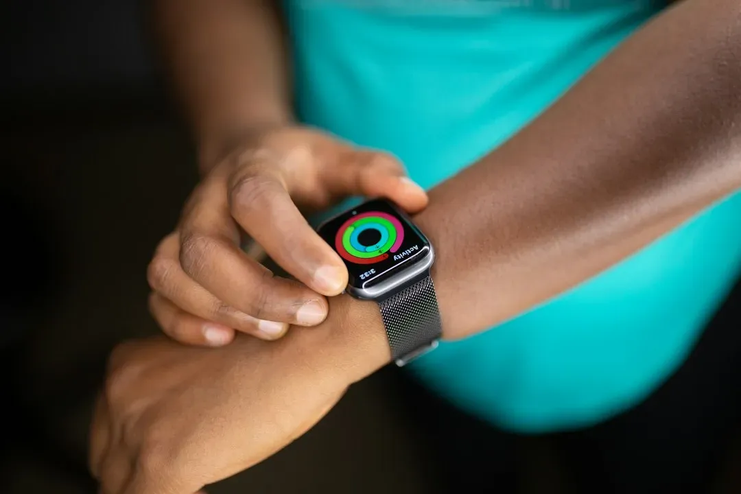
Looking inside without cutting is no longer confined to giant machines in hospitals. Over the last two decades, a quiet revolution has brought sensing closer to daily life. Wearable devices track heart rhythms, sleep patterns, physical activity, and sometimes even blood oxygen or continuous glucose levels, turning bodies into data streams that can be watched in real time.
On the medical edge, cardiac monitors that used to require bulky external boxes are now tiny patches or even implantable loop recorders, logging heart rhythm for months or years. Home ultrasound devices, while still limited, are starting to trickle into clinics and, in some places, even consumer hands. Smartwatches can flag irregular heart rhythms that might suggest atrial fibrillation, prompting people to seek evaluation earlier than they otherwise would have.
This everyday monitoring has clear benefits but also new tensions. It democratizes information – you no longer need a formal appointment to notice that your resting heart rate has been creeping up or your sleep quality is consistently poor. At the same time, it generates an avalanche of data that can cause anxiety, false alarms, and an understandable temptation to chase every small blip with medical testing.
In my view, this is one of the most emotional shifts of the entire story. Bodies used to be black boxes that you checked only when something felt wrong. Now many people carry a quiet medical lab on their wrist, watching themselves in a way that blends curiosity, reassurance, and sometimes an uncomfortable sense of never being fully “off the clock” from their health.
#7 Transforming Research Design: From Single Snapshots To Moving Pictures
Before non‑invasive imaging, much of biological and medical research worked like a photo album: a collection of still shots from different individuals at different times, stitched together into a guess about progression. You might compare one group of patients with early disease to another group with advanced disease and infer how things changed in between, without ever watching those changes unfold in the same person.
Once researchers could look inside the same individual repeatedly, everything shifted toward moving pictures instead of scattered photographs. Longitudinal imaging studies could follow the same heart, brain, or tumor over months or years, tracking subtle changes and how they related to symptoms, lifestyle, or treatments. That kind of within‑person tracking is far more powerful for understanding cause and effect.
Non‑invasive techniques have also allowed scientists to connect layers that were previously separate. For example, they can pair genetic data with serial scans to see how certain variants influence brain development or vascular plaque growth. Clinical trials can now use imaging markers as early indicators of whether a therapy is working, long before traditional outcomes like survival or symptom scores are fully known.
In practice, this has led to research that feels more like watching a documentary than flipping through strangers’ photos. It has also forced the field to confront new analytical challenges: enormous data sets, complex time series, and the need for advanced statistics and machine learning just to make sense of what we are now able to see.
#8 Engineering, Materials, And Machines: Looking Inside Things That Are Not Alive
Medicine grabs most of the attention, but the same core idea – seeing inside without cutting – has also transformed engineering and materials science. Non‑destructive testing techniques like industrial X‑ray, ultrasound, and advanced microscopy let researchers and manufacturers inspect aircraft wings, bridges, batteries, and microchips for hidden defects without destroying the part.
In aviation, for example, safety depends on catching tiny cracks and corrosion long before they cause catastrophic failures. High‑resolution imaging can reveal flaws deep within metal or composite structures, guiding repairs or replacements while keeping planes in service safely. Similarly, in the energy sector, imaging of pipelines and reactors can detect weak points that would be dangerous to assess by taking things apart in the usual way.
On the research side, watching materials deform, corrode, or age from the inside is a game changer. Scientists can subject samples to heat, stress, or chemical exposure while using imaging to see how internal structures respond in real time. That kind of insight feeds directly into better designs: lighter but stronger alloys, more durable batteries, and safer electronic components.
What I love about this parallel story is how similar the pattern is to medicine. First you get crude glimpses, then clearer images, then dynamic, real‑time views that change how you design and maintain the whole system. Whether it is a human joint or a jet engine blade, looking inside without cutting nudges every field toward prevention, prediction, and smarter intervention.
#9 Ethics, Privacy, And The Psychology Of Being So Visible
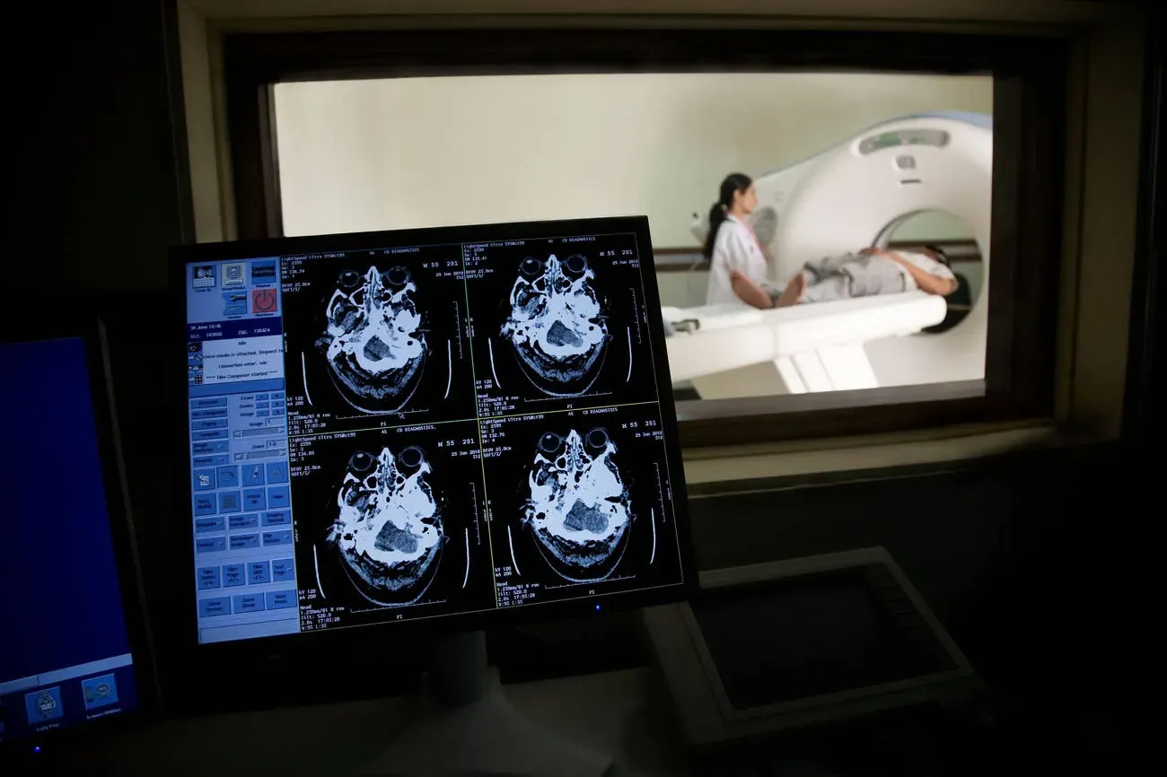
Seeing more is not automatically an uncomplicated good. Once researchers and clinicians , they also inherited gnarly ethical questions that are nowhere near settled. If a scan done for research accidentally reveals something concerning – a small aneurysm, a suspicious nodule – what exactly is the obligation to tell the subject, and who pays for the follow‑up?
Privacy is another thorny area. Internal images and biometric data are deeply personal. As imaging and sensors move into the cloud and into consumer devices, lines blur between medical information, wellness tracking, and corporate data collection. Laws and norms are still catching up, and people are rightly wary of a world where insurers, employers, or tech companies might access or infer intimate details from their internal “maps.”
On a more subtle level, there is the psychological impact of knowing so much about your own insides. For some, scans bring huge relief – a clean imaging result can quiet fears. For others, especially those prone to health anxiety, the possibility of finding small anomalies everywhere can be overwhelming. Medicine has had to learn a new kind of counseling: explaining not just what was found, but what it means to live with incidental findings that may never cause harm.
Summarizing some of the key tensions:
- How to balance the duty to inform with the risk of triggering needless fear.
- How to protect imaging data in a world of massive, often commercial, data flows.
- How to help people live with the knowledge that being “normal” on the inside often still looks a bit messy on a scan.
In many ways, these ethical and emotional questions are the shadow side of the visibility we worked so hard to achieve.
#10 Where We Go Next: My Take On A Future Of Radical Transparency
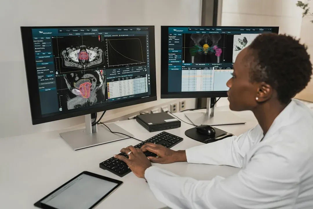
Looking back, the story of non‑invasive imaging feels like a slow but unstoppable march toward radical transparency. We started out blind, learned to glimpse bones, then organs, then flowing blood, firing neurons, and microscopic structures. With each step, outcomes improved, guesses shrank, and entire research agendas were rewritten. We now take for granted that a chest pain workup includes scans, that surgical plans are drawn on cross‑sectional images, and that brain research means watching living brains, not just dissecting silent ones.
In my opinion, the next phase will be less about inventing entirely new ways of looking and more about weaving everything together into coherent stories. We are drowning in data – images, waveforms, sensor logs – but still learning how to combine them into simple, trustworthy answers that help people make decisions. Artificial intelligence will almost certainly play a larger role here, flagging patterns that humans miss, but it will also raise tough questions about trust, bias, and who ultimately bears responsibility when an algorithm reads your insides.
I also suspect we are underestimating how much this deep visibility will change our culture. When you grow up in a world where scanning your brain or your genome is normal, your relationship to fate, responsibility, and risk looks different. You may feel more empowered, but also more exposed. It is tempting to imagine a future where everyone walks around with a detailed digital twin of their body, updated by periodic scans and wearables, used to simulate “what if” scenarios before choosing treatments or lifestyles.
That vision excites me and worries me in equal measure. We have already seen what happens when technologies that start as tools for care become engines for surveillance or profit. The challenge now is to protect the best parts of being able to look inside without cutting – the rescue of patients from needless suffering, the leap forward in understanding complex systems – while drawing firm lines around how, when, and why that power is used. In the end, the real question might be less about what we can see, and more about what kind of society we want to build now that our insides are no longer invisible. What would you want to know if you could see everything?