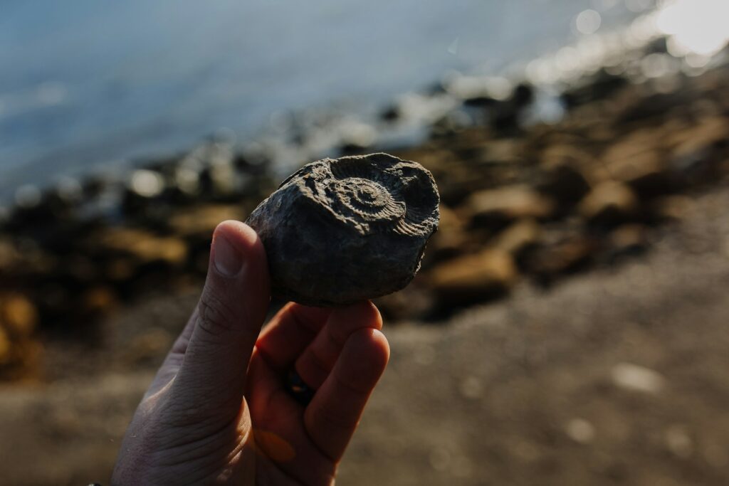
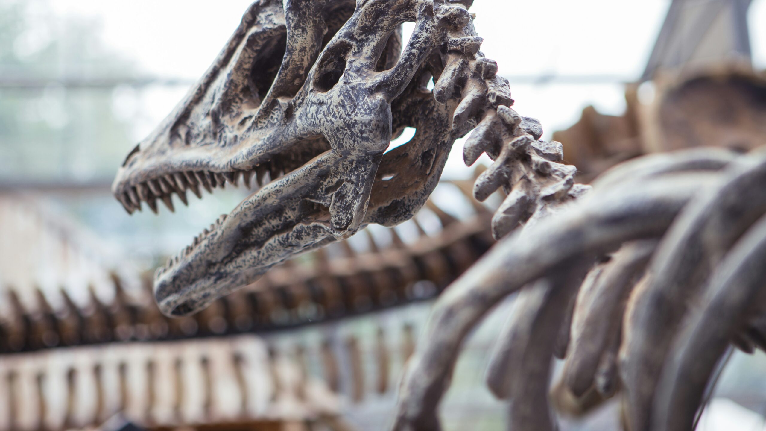
In the quiet halls of museums worldwide, ancient bones whisper stories of the past to those who know how to listen. Today, scientists are hearing these stories with unprecedented clarity thanks to revolutionary technological tools. The emergence of advanced imaging technologies, laser scanning systems, and robotic assistants has fundamentally transformed paleontology and archaeology, allowing researchers to extract information from fossils and ancient remains that would have been impossible just decades ago. From the internal structures of dinosaur skulls to the microscopic growth patterns in hominin teeth, modern technology is illuminating our understanding of ancient life in remarkable ways. This technological renaissance represents one of the most significant advances in the study of ancient remains since the development of radiocarbon dating in the 1940s, opening new windows into prehistoric worlds.
The Evolution of Paleontological Research Methods

The study of ancient bones has undergone a dramatic transformation since its earliest days. Traditional paleontological methods relied heavily on physical examination, measurement, and destructive sampling techniques that often damaged precious specimens. Researchers would physically section fossils to examine internal structures, losing information and potentially damaging unique specimens in the process. The limitations of these approaches created significant blind spots in our understanding of ancient life. By the mid-20th century, X-ray technology began offering non-destructive glimpses inside fossils, but resolution remained poor, and many structures remained invisible to researchers. The digital revolution of the late 20th and early 21st centuries marked a paradigm shift, introducing tools that could peer into fossils with microscopic precision while leaving specimens completely intact. This technological evolution has democratized research, allowing scientists to share perfect digital replicas of rare specimens with colleagues worldwide, dramatically accelerating the pace of discovery.
CT Scanning: Seeing Inside Ancient Remains
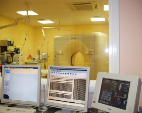
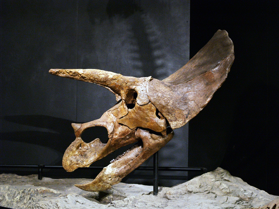
Computed tomography (CT) scanning has revolutionized how scientists study ancient bones by allowing them to visualize internal structures without damaging specimens. Unlike conventional X-rays that produce flat, two-dimensional images, CT scanners generate thousands of cross-sectional images that can be assembled into detailed three-dimensional reconstructions. This technology works by directing X-rays through a specimen from multiple angles while detectors measure the variation in X-ray attenuation, creating “slices” that reveal density differences within the object. Modern micro-CT scanners can achieve resolutions of just a few micrometers, allowing researchers to examine microscopic structures like blood vessel channels or growth lines in teeth. The non-destructive nature of CT scanning is particularly valuable for rare or delicate specimens that cannot be sectioned for traditional analysis. For example, CT scanning has allowed scientists to examine the brain cases of dinosaur skulls, revealing details about neural anatomy that were previously impossible to study.
Synchrotron Imaging: The Subatomic Fossil Detective
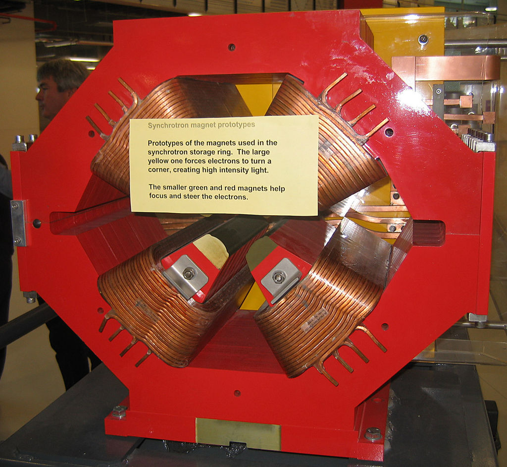
Synchrotron radiation has emerged as one of the most powerful tools for examining ancient remains at nearly atomic scales. These massive circular particle accelerators generate extremely bright X-rays billions of times more intense than those used in hospital CT scanners, allowing for unprecedented resolution and contrast sensitivity. Synchrotron imaging can differentiate tissues based on subtle chemical differences, revealing details invisible to conventional imaging techniques. At facilities like the European Synchrotron Radiation Facility in France or the Advanced Photon Source in the United States, paleontologists can detect fossilized soft tissues, examine microscopic growth patterns, and even identify chemical signatures that reveal ancient diets or environments. The technique has produced breakthrough discoveries, including the visualization of 195-million-year-old dinosaur embryos still inside their eggs and the identification of preserved protein fragments in 75-million-year-old dinosaur fossils. These capabilities come at a cost, however—synchrotron facilities are billion-dollar installations with limited availability, creating fierce competition for research time.
3D Laser Scanning: Capturing Surface Details
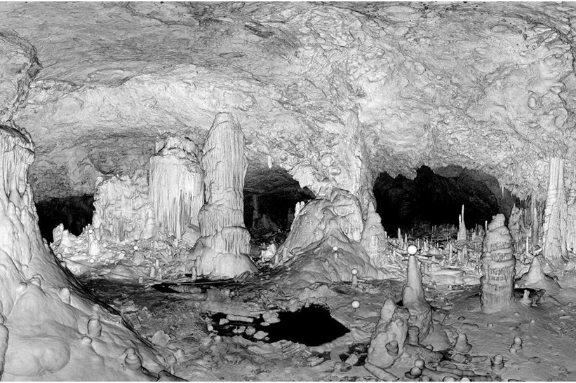
Three-dimensional laser scanning technologies have transformed how researchers document and analyze the external morphology of ancient bones. These systems project laser light onto specimen surfaces and measure the reflected light to create precise “point clouds” representing the object’s external geometry. Modern laser scanners can capture surface details with sub-millimeter accuracy, preserving information about tool marks, bite marks, pathologies, and subtle anatomical features. Unlike photography, which captures only what’s visible from specific angles, 3D scanning creates complete digital models that can be manipulated, measured, and analyzed from any perspective. This technology has proven particularly valuable for tracking taphonomic changes in bones—the alterations that occur during decomposition, fossilization, and geological processes. Portable laser scanners have also revolutionized fieldwork, allowing paleontologists and archaeologists to create precise digital records of specimens or entire excavation sites before removing fragile materials from their discovery contexts. The resulting digital models serve as permanent records that can be studied long after specimens have been collected or reburied.
Photogrammetry: Democratizing Digital Documentation
Photogrammetry has emerged as a remarkably accessible technology for creating detailed 3D models of ancient bones through sophisticated computational analysis of ordinary photographs. The technique requires no specialized equipment beyond a digital camera, making it extraordinarily cost-effective compared to laser scanning systems that can cost tens of thousands of dollars. Researchers capture dozens or hundreds of overlapping photographs of a specimen from different angles, then use specialized software to identify matching points across images and calculate the camera’s position for each photo. From this information, the software generates a 3D point cloud and textured mesh that accurately represents the specimen’s shape and appearance. The democratizing effect of photogrammetry has been profound, allowing researchers with limited resources to create and share high-quality 3D models of specimens. Archaeological projects increasingly use drone-mounted cameras to perform photogrammetry of entire excavation sites, creating comprehensive digital records that preserve spatial relationships between discovered remains. The technique’s main limitation is its inability to capture internal structures, making it complementary to rather than a replacement for CT scanning.
Robotic Assistance: Precision Handling and Analysis
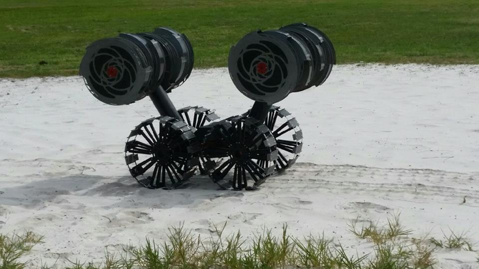
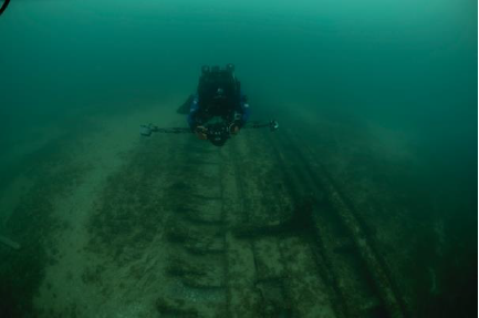
Robotic systems are increasingly finding applications in the study of ancient remains, offering advantages in precision, consistency, and the ability to work with fragile specimens. Custom-designed robotic arms equipped with force sensors can handle delicate fossils with precisely controlled movements, minimizing the risk of damage during examination or preparation. Some laboratories have developed automated systems that can extract microfossils from sediment samples or take standardized photographic series for photogrammetry with perfect consistency. Advanced robotics systems are also being developed to assist with field excavations, using artificial intelligence to identify bone fragments in complex matrices and extract them with minimal damage. Perhaps most impressive are the semi-autonomous robots designed for underwater archaeology, which can document shipwreck sites or submerged settlements at depths inaccessible to human divers. The Woods Hole Oceanographic Institution’s remotely operated vehicles have recovered human remains from historical shipwrecks at depths exceeding 4,000 meters, demonstrating how robots can extend archaeological research into previously inaccessible environments.
Digital Reconstruction: Bringing Fragments Together
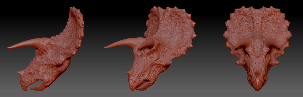
Digital reconstruction technologies have transformed how researchers work with incomplete or fragmented ancient remains, allowing virtual reassembly of specimens too delicate for physical handling. Using 3D models created through CT scanning or laser scanning, scientists can digitally manipulate bone fragments, testing different configurations to determine correct anatomical relationships. This approach offers significant advantages over physical reconstruction, as it allows researchers to easily undo unsuccessful attempts without risking damage to the original specimens. Software tools can automatically identify matching surfaces between fragments, suggest likely configurations, or even compensate for missing sections using morphological patterns from related species. These techniques have proven particularly valuable for analyzing remains affected by taphonomic distortion, where bones have been warped by geological processes over time. Digital reconstruction has enabled dramatic reassessments of controversial specimens like the Taung Child (Australopithecus africanus) and Homo floresiensis, allowing researchers to correct for postmortem distortion and reveal the true anatomical configurations of these important hominin fossils.
Artificial Intelligence in Ancient Bone Analysis

Artificial intelligence and machine learning are creating new possibilities for analyzing the vast datasets generated by modern imaging technologies. Neural networks trained on thousands of bone specimens can automatically identify species from fragmentary remains with accuracy sometimes exceeding that of human experts. This capability is particularly valuable for processing large collections of microfossils or fragmented remains from archaeological sites, where manual identification would be prohibitively time-consuming. AI systems are also being developed to detect subtle patterns invisible to human observers, such as microscopic cut marks that distinguish human butchery from animal tooth marks or weathering effects. Perhaps most promising are deep learning approaches that can identify patterns across thousands of specimens, revealing evolutionary relationships or anatomical correlations that might escape traditional comparative methods. The Allen Institute for AI’s Fossil Finder project exemplifies this approach, using computer vision algorithms to scan thousands of fossil images and identify specimens of interest that merit closer human examination. As these systems become more sophisticated, they increasingly serve as research partners rather than mere tools, suggesting new hypotheses and analytical approaches.
X-ray Fluorescence: Reading Chemical Signatures

X-ray fluorescence (XRF) technology has opened new avenues for studying the chemical composition of ancient bones without destructive sampling. This technique works by directing X-rays at a specimen, causing atoms in the material to emit secondary X-rays with energies characteristic of specific elements. Portable XRF analyzers now allow researchers to perform elemental analysis in the field or museum, identifying the presence and concentration of elements from calcium and phosphorus to trace metals like strontium, lead, or mercury. These chemical signatures can reveal crucial information about an individual’s diet, habitat, or exposure to environmental toxins. Strontium isotope ratios, for example, vary geographically based on local geology, allowing researchers to determine if an individual migrated during their lifetime by comparing dental enamel (formed in childhood) with bone (which continuously remodels). XRF analysis has identified lead poisoning in ancient Roman remains, mercury treatments in medieval burials, and arsenic exposure in prehistoric mining communities. When combined with spatial mapping techniques, XRF can create detailed elemental maps showing the distribution of specific elements across a specimen, revealing patterns of mineral replacement during fossilization or localized exposure to environmental contaminants.
Biomechanical Modeling: Understanding Ancient Movement
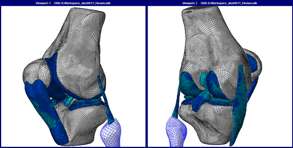
Advanced computational techniques are allowing researchers to move beyond static anatomical studies to explore how ancient creatures actually moved and functioned. Finite element analysis (FEA), originally developed for engineering applications, uses digital models to simulate how structures respond to physical forces, revealing how bones would have performed under different mechanical stresses. This approach allows researchers to test hypotheses about extinct animals’ behaviors by simulating activities like biting, running, or flying and analyzing the resulting stress patterns. Multibody dynamics analysis takes this further by modeling entire musculoskeletal systems, simulating coordinated movement of multiple bones connected by virtual muscles and tendons. These techniques have resolved longstanding debates about dinosaur locomotion, demonstrating that Tyrannosaurus rex likely could not run at high speeds despite its fearsome reputation, as its skeleton would have experienced catastrophic stress failures. Similar approaches have clarified the locomotive capabilities of early hominins like Australopithecus afarensis (represented by the famous “Lucy” skeleton), demonstrating a unique bipedal gait different from modern humans. As computational power increases, these simulations grow increasingly sophisticated, incorporating soft tissue properties and neurological control systems to create more accurate models of ancient movement.
Ancient DNA Analysis: The Molecular Fossil Record

Revolutionary advances in ancient DNA extraction and sequencing technologies have created an entirely new dimension in the study of ancient remains. While not strictly imaging technologies, these molecular approaches complement morphological studies by revealing genetic relationships invisible to visual inspection. Next-generation sequencing platforms can recover and analyze fragmentary DNA molecules preserved in bones thousands or even hundreds of thousands of years old, reconstructing partial or complete genomes of extinct species. These methods have completely transformed our understanding of human evolution by revealing interbreeding between Homo sapiens and Neanderthals, identifying previously unknown hominin groups like the Denisovans, and tracing population movements across continents. In paleontology, ancient DNA has resolved longstanding debates about evolutionary relationships, demonstrating that birds are the direct descendants of theropod dinosaurs and clarifying the relationship between woolly mammoths and modern elephants. The integration of genetic data with morphological studies has revealed cases where similar anatomical features evolved independently in unrelated lineages (convergent evolution) and identified cryptic species that appear physically similar but represent distinct genetic lineages. As extraction and analysis methods improve, researchers continue pushing back the temporal limits of ancient DNA recovery, with the current record held by mammoth DNA recovered from permafrost deposits 1.2 million years old.
Augmented and Virtual Reality in Paleontological Research
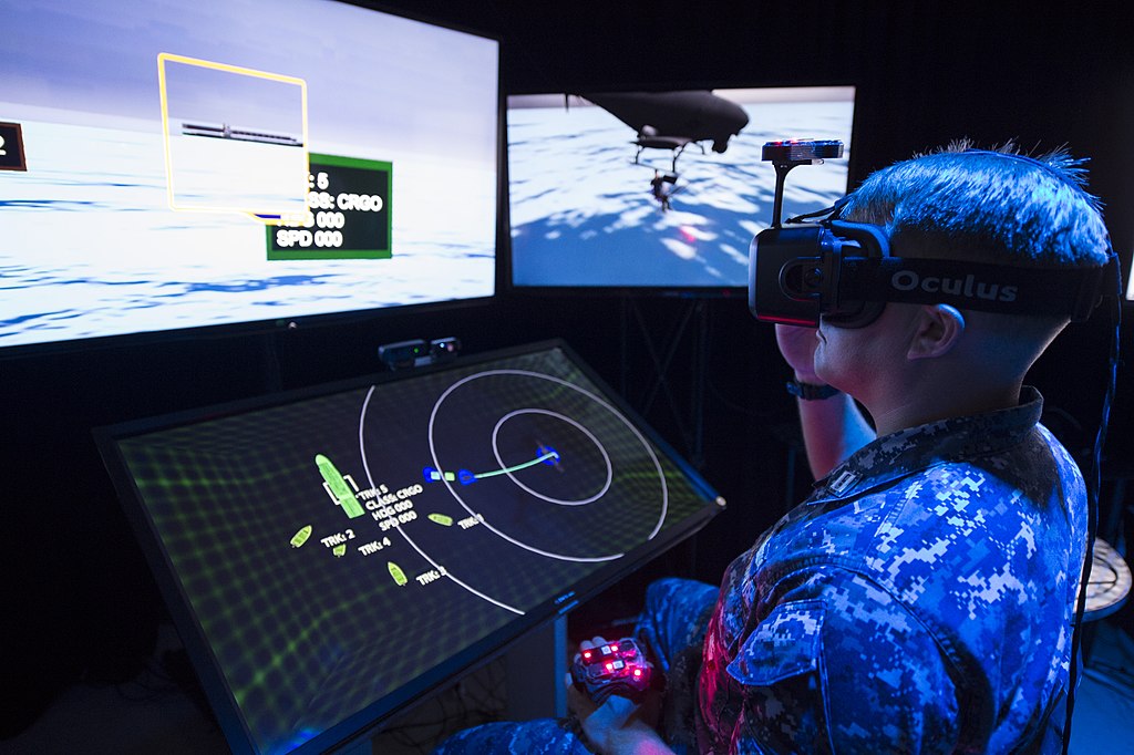
Augmented and virtual reality technologies are creating new ways for researchers to interact with digital models of ancient remains, offering intuitive interfaces for examining complex three-dimensional structures. VR systems allow scientists to manipulate virtual specimens at any scale, walking around life-sized dinosaur reconstructions or zooming in to examine microscopic features of teeth or bone microstructure. These immersive environments are particularly valuable for collaborative research, allowing specialists in different locations to examine and discuss the same virtual specimens in real-time shared spaces. Augmented reality applications overlay digital information onto physical specimens or excavation sites, helping researchers visualize relationships between fragmented remains or providing contextual information about discovery locations. Museums and research institutions increasingly use these technologies to enhance access to rare specimens, creating virtual repositories that can be accessed remotely by researchers worldwide. The Smithsonian Institution’s Digitization Program Office exemplifies this approach, creating detailed 3D models of significant specimens that can be examined through VR interfaces by researchers unable to visit the physical collections. These technologies are transforming not just research practices but also educational approaches, allowing students to virtually handle and examine specimens that would be too valuable or fragile for classroom use.
Ethical Considerations in High-Tech Research
The application of advanced technologies to the study of ancient human remains raises important ethical questions that researchers must navigate thoughtfully. While non-destructive imaging techniques offer clear benefits by preserving physical specimens, the creation of detailed digital models introduces new complexities around data ownership, access rights, and appropriate representation of culturally sensitive materials. Indigenous communities worldwide have raised legitimate concerns about the digitization of ancestral remains, questioning whether virtual reconstructions might violate cultural prohibitions against displaying or studying certain remains. These considerations have led to important conversations about informed consent protocols for digital imaging, appropriate limitations on visual representations, and community involvement in research decisions. The development of ethical frameworks for digital paleontology and bioarchaeology represents an evolving area of practice, with many institutions now implementing consultation processes and collaborative research models. Some projects have pioneered innovative approaches, such as the “digital repatriation” of virtual models to source communities while physical specimens remain in museum collections, or the creation of tiered access systems that respect cultural protocols while enabling scientific research. These ethical frameworks recognize that technological capabilities must be guided by respect for the communities connected to the remains being studied and acknowledgment of historical injustices in scientific collection practices.
Future Horizons: Emerging Technologies in Ancient Bone Research
The technological frontier in ancient bone research continues advancing rapidly, with several promising developments on the horizon. Hyperspectral imaging systems, which capture data across hundreds of spectral bands rather than just visible light, are beginning to reveal previously invisible organic residues on ancient remains, identifying preserved proteins, fats, and plant materials. Quantum computing applications are being developed that may dramatically accelerate the processing of massive datasets from high-resolution imaging, potentially allowing real-time analysis of synchrotron data or instantaneous comparison of specimens against global databases. Miniaturization of advanced imaging technologies is creating new possibilities for field application, with portable CT scanners and synchrotron-like X-ray sources that could revolutionize on-site analysis of newly discovered remains. Perhaps most exciting is the integration of these various technologies into comprehensive analytical platforms that combine morphological, chemical, genetic, and contextual data to provide holistic views of ancient remains. As these technologies mature, they promise to continue transforming our understanding of ancient life, revealing new details about the creatures that inhabited our planet millions of years ago and the evolutionary processes that shaped our own species. The coming decades will likely witness discoveries that fundamentally reshape our understanding of life’s history on Earth, driven by these remarkable technological tools.
Conclusion: The Technological Renaissance in Paleontology

The integration of advanced imaging, digital reconstruction, and computational analysis has fundamentally transformed how scientists study ancient bones, creating a renaissance in paleontological and archaeological research. These technologies have removed longstanding barriers to analysis, allowing non-destructive examination of previously inaccessible structures, precise quantification of morphological features, and sophisticated testing of functional hypotheses. The digital nature of modern research has democratized access to rare specimens through shareable 3D models while facilitating global collaboration among specialists.