Imagine being able to look inside a 70-million-year-old skull without ever touching it. No chisel, no drill, no risk of shattering something irreplaceable. That idea, which would have seemed like science fiction just decades ago, is now everyday reality for paleontologists around the world. The tools they are working with today are so advanced that they are rewriting what you thought you knew about some of the most iconic creatures to ever walk the Earth.
We are living through what might honestly be described as a golden age of dinosaur science. Not because we are finding more bones, though that is happening too, but because new imaging technologies are letting researchers peer inside fossils in ways never before possible. The results are surprising, thrilling, and sometimes downright shocking. Let’s dive in.
The CT Scanner: From Hospital to Fossil Lab
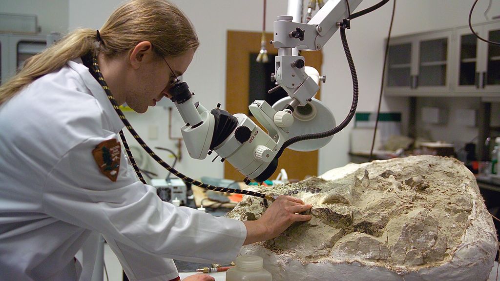
In the past few decades, Computed Tomography (CT) scanning of fossil material has become the tool of choice for most paleontologists wanting a non-destructive way to extract fragile fossils from their encasing matrix or to investigate their internal anatomy. Think about that for a second. The same basic technology your doctor uses to check for a broken rib is now being used to reveal the hidden anatomy of a Tyrannosaurus rex. It is one of those cross-disciplinary collisions that makes science so exciting.
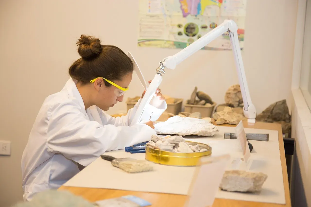
Traditionally, researchers used destructive thin sectioning to reveal interior structures, which totally destroys the fossils. With the application of non-destructive 3D imaging techniques like CT scanning and synchrotron radiation scanning, paleontologists can now observe and interact with previously hidden structures without causing any damage. For a field built around protecting rare, one-of-a-kind specimens, this shift is nothing short of revolutionary. You can now examine what would have previously required permanent destruction of an object that took millions of years to form.
Synchrotron Scanning: The Most Powerful Imaging Tool in Paleontology
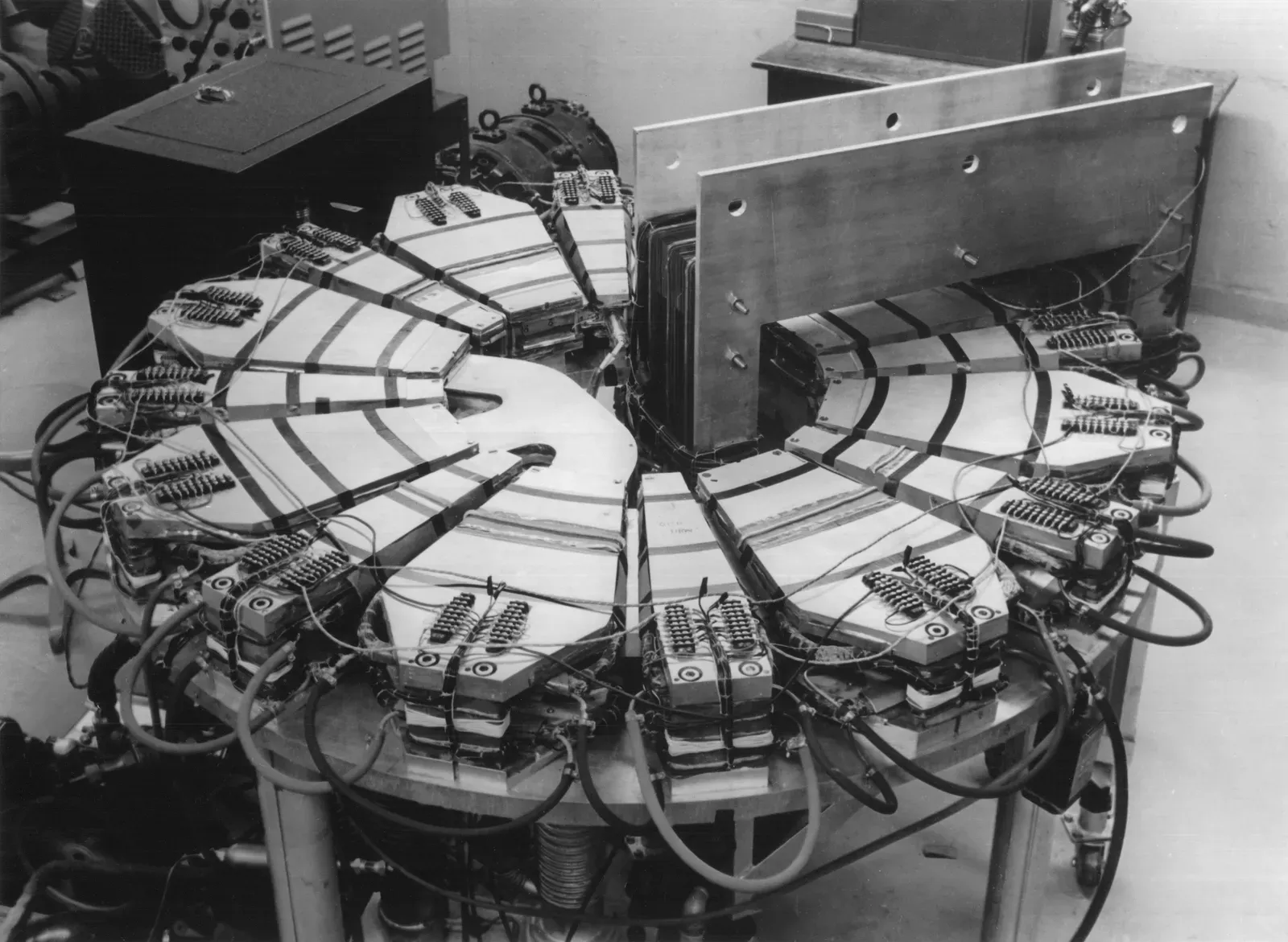
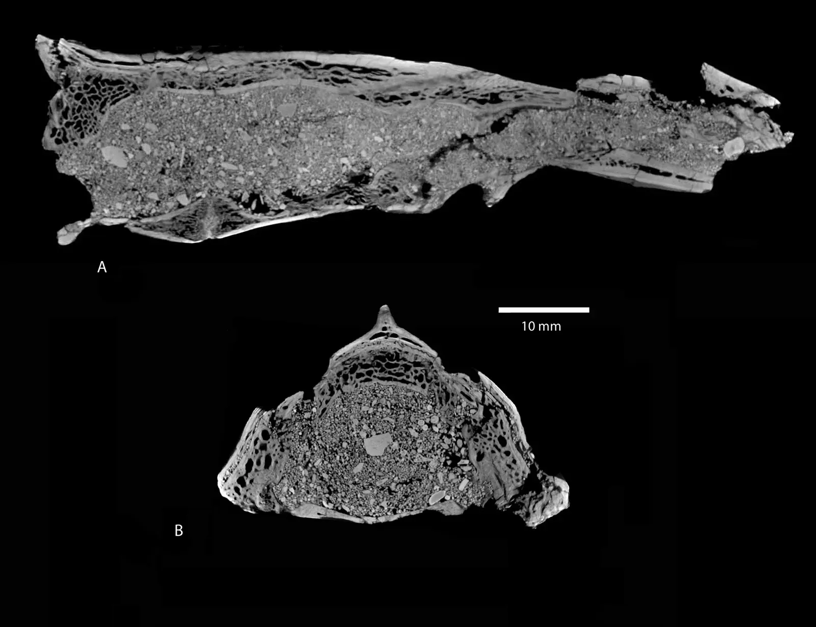
Here is the thing about regular CT scanners. They are good, but they have limits. Synchrotron scanning takes things to a completely different level. Researchers at Australia’s Synchrotron used the Imaging and Medical beamline to acquire detailed images of fossils, including a shin bone belonging to one of Australia’s largest known carnivorous dinosaurs. The sample challenged the very limits of the beamline’s physical capability, requiring the highest energy X-rays of the brightest X-ray source in the southern hemisphere to peer inside the bone.
Synchrotron CT scanning is genuinely exciting because, if it comes into more widespread use, it has the potential to reveal so much information about dinosaurs, their skulls, and the features of their bones. Synchrotron sources take advantage of highly brilliant and coherent X-rays, which enable fast data acquisition and enhanced image contrast for biological samples owing to the exploitation of phase contrast. In plain terms: you get richer, sharper, more detailed images than almost anything else available to science today.
Unlocking Dinosaur Brains and Neuroanatomy
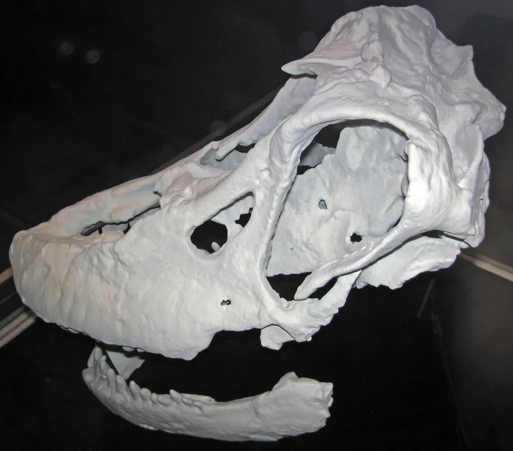
You have probably never stopped to wonder what a dinosaur’s brain actually looked like from the inside. Honestly, neither had most scientists until the imaging tools to answer that question finally arrived. A true turning point came with the efforts of Timothy Rowe, who in the 1990s helped create a laboratory at the University of Texas at Austin dedicated to producing high-resolution CT data of fossils. This non-destructive method bears the inherent potential to produce and study anatomically detailed digital endocasts with relative ease.
Today, most centers of paleontological research include a CT laboratory, with endocast construction being standard operating procedure. Digital approaches, now including X-ray synchrotron and neutron X-ray CT, also facilitate the objective retrodeformation of taphonomic damage. In other words, scientists can now digitally “undo” the crushing and warping that happens to fossils over millions of years, essentially restoring a skull to its original shape on a computer screen. Because fossils usually do not preserve molecular or behavioral information, paleontologists focus mainly on morphology, which includes not only exterior features but also interior structures like brain endocasts and inner ears.
Artificial Intelligence: Turbocharging Fossil Analysis
Here is where things get almost startling. Imaging technology is powerful on its own, but when you combine it with artificial intelligence, the pace of discovery accelerates dramatically. Researchers have developed a new AI algorithm that uses high-resolution CT imaging and deep learning models to scan and evaluate dinosaur fossils. The algorithm was tested on CT scans of Protoceratops dinosaur fossils and achieved a high accuracy of around 97 percent in segmenting fossils from rock, significantly reducing the time required for this process.
While the feature model did not perform as accurately as scientists in all tasks, the segmentation models worked smoothly and did so in record time, segmenting each slice in seconds. Manually segmenting the same piece took minutes or even hours in some cases. That compression of time is enormous. What once cost a researcher weeks of painstaking manual labor can now be accomplished in a fraction of the time. This workflow has the capacity to revolutionize the use of deep learning to significantly reduce the processing time of such data and boost the availability of segmented CT-scanned fossil material for future research outputs.
Drones, Photogrammetry, and the Art of Finding Fossils First
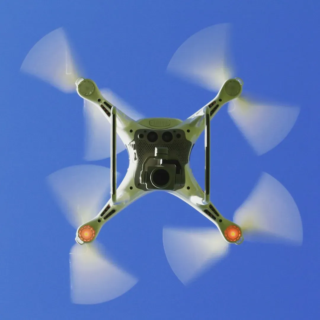
You might be wondering: what does a drone have to do with dinosaur anatomy? More than you would think. Before any scan can be performed, scientists need to find the fossils. Researchers used drones to capture around a thousand high-resolution images of a key fossil site in Dinosaur Provincial Park. These images were processed through a technique called structure-from-motion photogrammetry, which allowed the team to construct a precise 3D model of the terrain geolocated with GPS coordinates measured in the field.
Studies have revealed that a long-relied-upon rock boundary at Dinosaur Provincial Park fluctuates in elevation by as much as 12 metres over short distances, introducing a level of uncertainty that could alter interpretations of when different species lived. As one researcher noted, if the reference point itself varies significantly, then estimates of individual fossil ages could be off by a considerable margin. This is a stunning finding. Drone-assisted photogrammetry is not just helping find fossils, it is correcting decades of assumptions about when those fossils actually lived. The application of remote sensing and drone imaging to help narrow down the best areas to prospect, combined with three-dimensional scanning and artificial intelligence to help identify problematic fossils, could revolutionize the entire field in the future.
3D Printing and the Democratization of Dinosaur Science
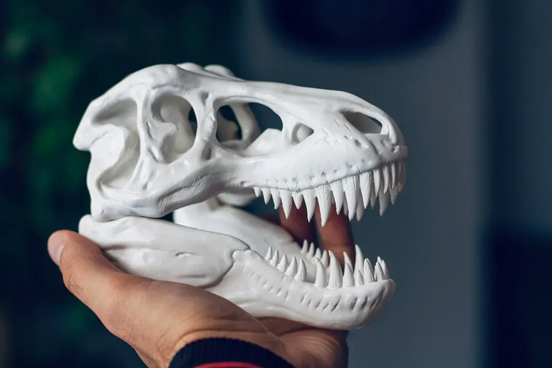
Once a fossil has been scanned, something incredible becomes possible. You can share it with the entire world. Once someone has completed a CT scan and produced a digital replica of a skull, that data can be easily shared with anyone around the world. Rather than traveling across the globe to examine a specimen in person, you can literally transfer the file to someone, making data availability far better and giving researchers access to specimens from all corners of the planet.
The discovery of Taurovenator in Argentina became a prime example of the integration of paleontology with emerging 3D technologies. Researchers used 3D scanning and printing techniques to digitally reconstruct and physically replicate its skeleton, thereby advancing research, preservation, and public science communication. Museums can print replicas. Students can hold a model of a T. rex jaw in their hands. 3D scanning is a far less invasive reproduction technique than casting and molding, meaning the original fossils can be more easily protected and conserved. The digital models themselves offer another form of conservation and archiving. Paleontologists no longer have to travel to the site of the fossils to research them; they can simply access detailed 3D models online.
New Species and Shocking Reclassifications Thanks to Imaging
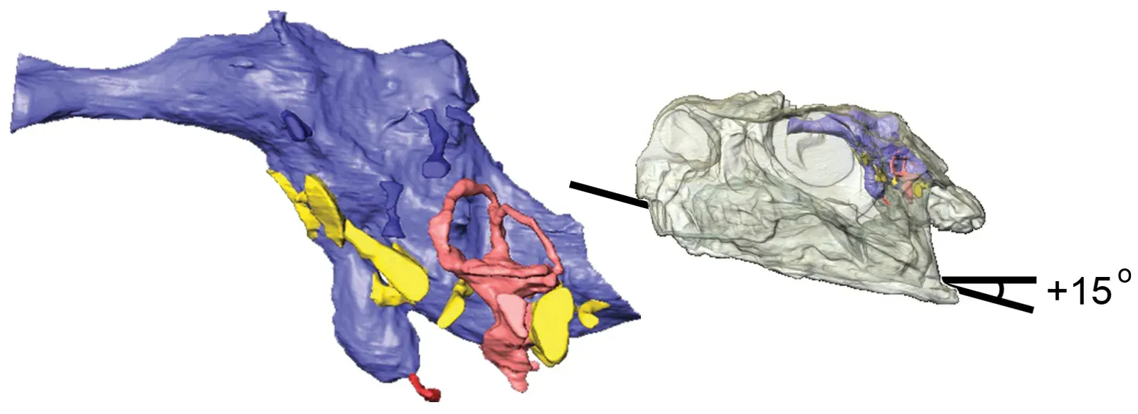
Perhaps the most jaw-dropping consequence of all this new imaging technology is what it is doing to our actual understanding of which dinosaurs are which. It is hard to say for sure just how many “discoveries” will ultimately upend the existing family tree, but the early signs are extraordinary. In one landmark case involving the “Dueling Dinosaur” fossil, researchers knew they needed to use the most cutting-edge technology to see inside the skull and look at bones that could not be seen with the naked eye. They took the skull to several CT and synchrotron scanning facilities across the United States to peer inside and reconstruct the internal anatomy of the tyrannosaur skull.
What the scans revealed was that the unique pattern of sinuses, the way the bones articulate in the skull, and the pattern of cranial nerves most closely matched the morphology of the skull known as Nanotyrannus lancensis, confirming a long-held suspicion among some paleontologists that it was not simply a teenage T. rex. Around 50 new dinosaurs are named each year and are discovered from across the globe, and the rate of new dinosaur discovery shows no signs of slowing down. Many of those discoveries and reclassifications are now being driven not by new digs, but by new scans of fossils that have been sitting in museum drawers for decades.
Conclusion: A New Era of Ancient Discovery
What is happening in paleontology right now is genuinely thrilling. New fossil finds and new methods, including modern computer imaging and simulation techniques, have changed our perception of animals that have been extinct for the last 66 million years or more. The creatures we thought we understood are revealing new secrets almost constantly, simply because we finally have the tools to ask better questions.
Think about it this way: the fossils have not changed. They have been sitting in the rock, in museum collections, waiting patiently. What has changed is your ability, and science’s ability, to actually see them. New generation CT scanners are proving to be powerful tools for the non-destructive imaging of large dinosaur fossils, with superior image quality and additional spectral information enhancing our understanding of fossil internal structure and composition, paving the way for new discoveries in paleontology. Every new scan, every AI-assisted analysis, every drone pass over a remote hillside carries the potential to change everything we thought we knew.
Somewhere out there, the next earth-shattering dinosaur discovery might not be hiding in the ground at all. It might already be sitting on a museum shelf, waiting for the right technology to finally reveal its secrets. What would you discover if you could look inside a 100-million-year-old skull?