Researchers unveiled Antscan, the world’s first comprehensive digital library of high-resolution 3D models capturing nearly 800 ant species across 212 genera worldwide.
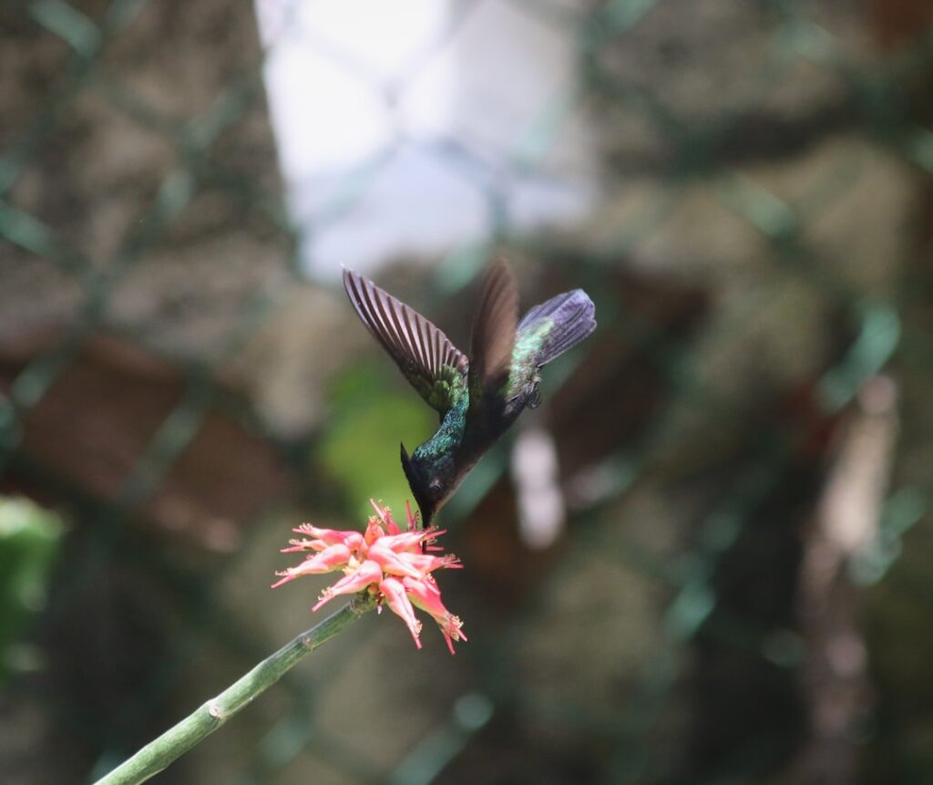
A High-Speed Revolution in Insect Imaging
A High-Speed Revolution in Insect Imaging (Image Credits: Imgs.mongabay.com)
Teams at the Karlsruhe Institute of Technology and the Okinawa Institute of Science and Technology pushed boundaries with synchrotron X-ray microtomography. This method, powered by a particle accelerator, generated intense X-ray beams to penetrate ant specimens in seconds. Robotic arms swapped samples every 30 seconds, enabling the scan of over 2,000 specimens in just one week – a feat that would have taken six years using traditional lab equipment.
Artificial intelligence then processed the raw data through platforms like Biomedisa. Neural networks segmented bodies, generated surface meshes, and reduced file sizes by 70 percent. The result offered pixel resolutions down to 1.22 micrometers, far surpassing conventional microscopy. Julian Katzke, the study’s first author, highlighted the efficiency: “With our setup, we scanned 2,000 specimens in a single week.”
Capturing the Breadth of Ant Biodiversity
The library documented 792 species from 14 of 16 ant subfamilies, representing more than half of all living genera. Workers dominated the collection at 1,671 specimens, followed by 291 queens and 220 males. Sources included museum collections, partner institutions, and experts globally, ensuring broad phylogenetic coverage.
Genera like Camponotus, Pheidole, and Strumigenys received dense sampling, with 37 to 39 species each. The dataset paired 3D phenotypes with genomic data for 186 species. All raw files and interactive viewers became freely available via the Antscan website, under a Creative Commons license.
Revealing Hidden Internal Worlds
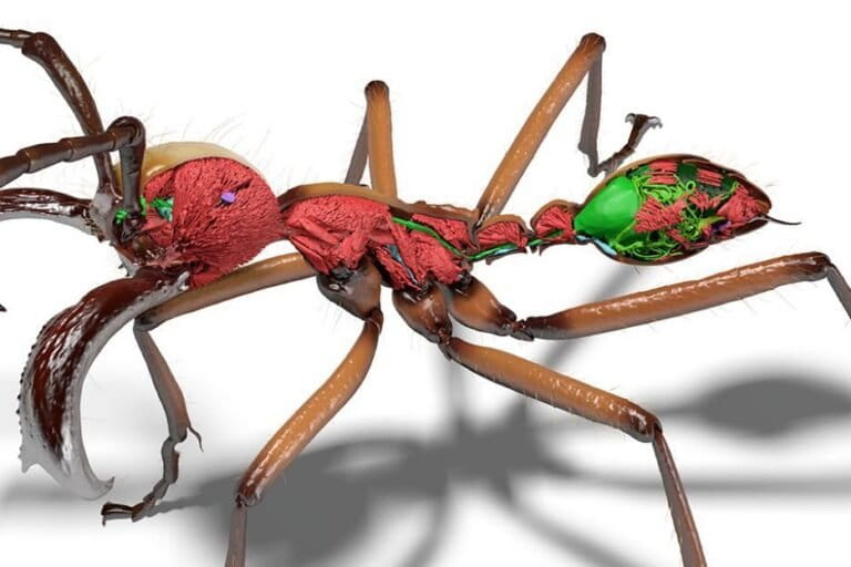
These scans pierced beyond exoskeletons to expose muscles, nervous systems, digestive tracts, and stingers at micrometer scales. Phase-contrast imaging highlighted soft tissues without staining, blending absorption data for comprehensive views. Such detail enabled novel analyses, like cuticle-to-body volume ratios across hundreds of species.
- Microscopic musculature patterns that inform locomotion studies.
- Nervous system layouts for behavioral research.
- Internal organ variations tied to ecological roles.
- Stinger and mandible structures for predatory insights.
- Exoskeleton thickness linked to colony dynamics.
Evan Economo, co-senior author from OIST, noted the accessibility: “We make it accessible to the whole world what otherwise would have been locked in a museum somewhere.”
Boosting Conservation Through Digital Archives
Ants, as ecosystem engineers, influence soil health, seed dispersal, and pest control across habitats. Yet, threats like habitat loss challenge their study. Antscan accelerated taxonomy and evolutionary research, integrating 3D data with genomics from prior OIST projects.
A 2025 study using early scans confirmed thicker cuticles correlated with smaller colonies, validating long-held hypotheses. The open platform invited citizen scientists, educators, and artists to explore. Economo envisioned broader impact: “This work moves us further into the big data era of capturing, analyzing, and sharing organismal shape and form.”
Key Takeaways
- Antscan digitized 792 species in 3D, covering over 50% of ant genera for global research.
- Synchrotron tech scanned 2,000+ specimens weekly, slashing digitization timelines.
- Open access empowers conservation, education, and innovative applications worldwide.
This blueprint promises scalable digitization for other invertebrates, potentially building a planetary biodiversity vault. As ants face environmental pressures, such resources will prove invaluable for protection strategies. What do you think about this leap in wildlife documentation? Tell us in the comments.