
Imagine a surgeon in 1950, scalpel in hand, staring at an organ he’s just been trained to call “useless.” He removes it without a second thought. Decades later, a researcher in a lab coat discovers that same organ was quietly protecting the immune system, cushioning a joint, or regulating hormones the whole time. This isn’t a hypothetical – it happened, over and over, across nearly every corner of the human body.
For most of modern medical history, the rule was simple: if a body part’s job wasn’t obvious under a microscope, it was probably evolutionary junk, and junk gets cut. Surgeons removed appendixes, tonsils, glands, and even chunks of cartilage on that assumption alone. It turns out doctors were wrong far more often than most people realize – and the science correcting them is stranger, and more unsettling, than you’d expect. Here’s what thirteen “pointless” body parts were actually doing all along.
#1 – The Appendix
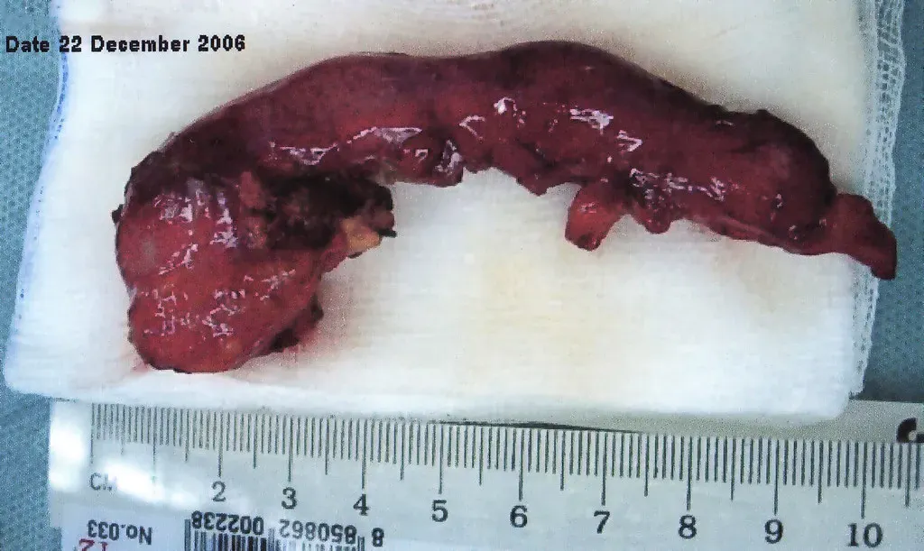
For more than a century, the appendix was treated as biological trash – a dead-end pouch hanging off the large intestine with no real job. Appendectomies became one of the most routinely performed surgeries on Earth, often done the moment inflammation showed up on a scan, no questions asked.
Then microbiome researchers actually looked closer. The appendix functions like a “safe house” for beneficial gut bacteria, sealed off by resilient biofilms that shield microbes from disruption. It’s a reservoir that can restock your gut’s bacterial population after an infection or a rough course of antibiotics wipes it out.
It’s also doing immune work most people never hear about. The appendix is packed with lymphoid tissue and is a major production site for immunoglobulin A, an antibody that keeps your intestinal flora balanced and in check.
Remove it, and the consequences are measurable. Studies link appendectomy to lower gut microbial diversity and a higher risk of several downstream conditions. Some researchers have even connected it to elevated odds of type 2 diabetes and Parkinson’s disease later in life – a shockingly high price for what was once called a spare part.
#2 – The Tonsils
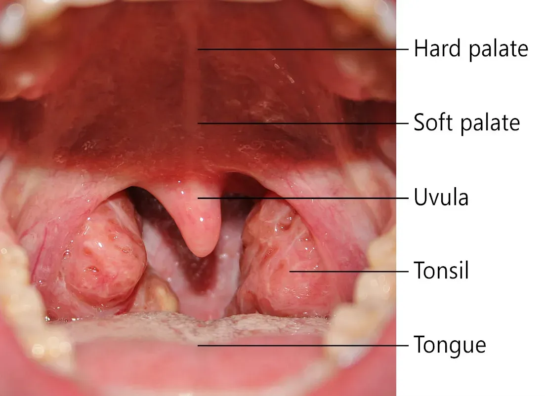
Tonsillectomies used to be a childhood rite of passage. Sore throat, recurring strep, a little snoring – it almost didn’t matter what the symptom was. Through the mid-20th century, surgeons pulled tonsils out by the millions, treating them as little more than troublesome scar tissue.
But the tonsils sit at the literal front gate of your immune system. They’re positioned right at the entrance of the throat, sampling everything you breathe, eat, and drink before it can travel further into your body.
Most people don’t realize tonsils are immune sentinels, not spare parts. They’re loaded with lymphocytes built to intercept invaders before an infection ever takes hold, and they help train the immune system – especially in early childhood, when the body is still learning what to fight.
Removing them doesn’t cripple your immunity, but it does strip away a layer of defense that took millions of years to build. Enough pediatric ENT specialists now consider routine tonsillectomy for mild, infrequent infections one of the most overused surgeries in medicine that the topic still sparks arguments in parenting forums everywhere.
Fast Facts
- Two lymphoid masses positioned at the throat’s entrance, screening food and air
- Packed with lymphocytes that intercept pathogens before they spread further
- Most active during early childhood, a key window for immune training
- Historically removed by the millions as a default fix for minor throat issues
#3 – The Adenoids
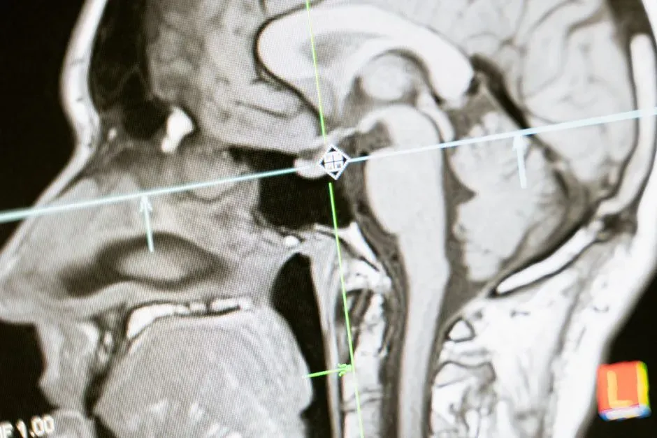
Tucked behind the nose and invisible without special equipment, the adenoids got even less respect than the tonsils. For decades, doctors removed them almost automatically whenever a child had chronic ear infections, breathing trouble, or snoring – often in the same surgery as a tonsillectomy, as a two-for-one.
Here’s the twist nobody explained to worried parents: adenoids are part of the same immune network as the tonsils, forming a section of what’s called Waldeyer’s ring – a circle of lymphoid tissue guarding the entrance to the respiratory and digestive tracts. In young children, whose immune systems are still under construction, this tissue plays an outsized role in building lifelong immunity.
The strange part is that this tissue naturally shrinks with age, which is exactly why removal seemed harmless for so long – doctors figured the body was already discarding something it didn’t need. But timing matters more than anyone realized. Pull adenoid tissue too early, before a child’s immune system has had a chance to “sample” enough pathogens through it, and you may short-circuit part of its natural education.
Most cases today only call for surgery when breathing or hearing is seriously affected – not as a routine, preventive default the way it once was.
#4 – The Thymus Gland
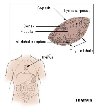
This is arguably the most dramatic reversal on the entire list. In the early 20th century, physicians became convinced that an enlarged thymus gland in infants was a ticking time bomb – a supposed condition called status thymicolymphaticus, the belief that an oversized thymus could compress the airway and cause sudden infant death.
Doctors began irradiating enlarged thymus glands in infants, with surgical removal as the fallback option. Thousands of children eventually received radiation treatment to prevent this “condition,” and some physicians even pushed for prophylactic irradiation on every newborn. There was just one problem.
The “enlarged” thymus was completely normal. The cadavers anatomists had used to define a “normal” thymus size mostly came from impoverished patients who’d died of chronic, stressful illnesses – and chronic stress is known to shrink the thymus. The benchmark itself was broken from the start.
In reality, the thymus is essential – it’s where T-cells mature and learn to recognize threats, forming the backbone of your adaptive immune system. Decades later, survivors of thymic irradiation still carry a measurably higher risk of thyroid cancer, showing up as far out as 58 years after treatment.
#5 – The Gallbladder
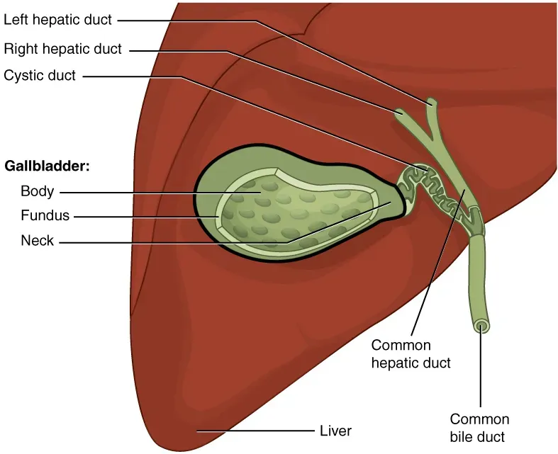
For years, the gallbladder was treated as an optional accessory to the liver – a small storage sac that wasn’t strictly necessary. Cholecystectomy remains one of the most common abdominal surgeries performed today, often for gallstones that cause pain but no lasting organ damage.
Here’s what tends to get glossed over in that pre-op conversation: the gallbladder doesn’t just passively store bile. It concentrates bile between meals and releases a precise, timed dose the moment fatty food hits the small intestine, letting your body break down fat in one coordinated burst.
Without it, bile trickles continuously from the liver instead of arriving on demand, and that steady drip can throw digestion out of rhythm.
Patients who’ve had their gallbladder removed often report years of unpredictable digestion afterward – bloating, diarrhea after fatty meals, and reduced absorption of fat-soluble vitamins A, D, E, and K. Surgeons will say the body “adapts” over time, and for most people it genuinely does. But plenty of gastroenterologists argue too many gallbladders get removed for stones that would have never triggered a real emergency in the first place.
#6 – The Spleen
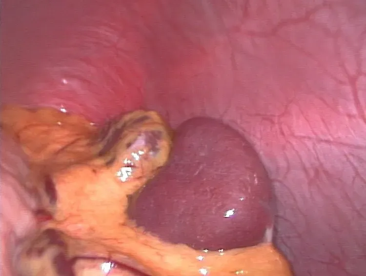
The spleen has long been the organ doctors were quickest to sacrifice, especially after trauma. A ruptured spleen from a car accident or a hard hit in sports used to mean immediate, total removal – better safe than risk internal bleeding, the logic went.
What surgeons underestimated is how much the spleen quietly does behind the scenes. It filters old and damaged red blood cells out of circulation, stores a reserve of platelets and white blood cells, and helps fight off certain bacterial infections – particularly encapsulated bacteria like the ones behind pneumonia and meningitis.
People who’ve had a splenectomy face a lifelong elevated risk of severe, sudden infections – a condition serious enough to have its own name: overwhelming post-splenectomy infection (OPSI). Because of this risk, patients without a spleen are typically required to get extra vaccinations, and some carry emergency antibiotics for the rest of their lives.
Modern trauma surgery has shifted hard toward spleen-preserving techniques whenever possible – a quiet admission that the old “remove it and move on” approach came with consequences nobody fully appreciated at the time.
#7 – Nasal Turbinates
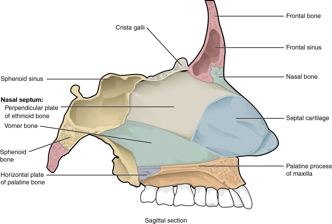
If you’ve never heard of turbinates, you’re not alone – but ENT surgeons have quietly performed turbinate reduction surgeries on chronically congested patients for decades, treating these small nasal structures as tissue standing in the way of easier breathing.
Each side of your nasal cavity has three turbinates – inferior, middle, and superior – bony shelves covered in mucosa that warm, humidify, and filter every breath you take. Remove too much of this tissue, and the fallout can be far worse than the congestion it was meant to fix.
Patients can end up with a condition called “empty nose syndrome.” It’s a bizarre, rare cluster of symptoms where you feel like you can’t get enough air, even though your nasal cavity is wide open – sometimes too open, with air rushing straight to the back of your throat. The nose looks physically fine on exam, yet patients feel chronically suffocated.
By 1990, the literature already showed that removing part or all of the lower turbinates wasn’t appropriate. Yet the surgery is still performed today, often without patients being warned this risk exists at all.
Worth Knowing
- Three turbinates line each nostril: inferior, middle, and superior
- Their job is to warm, humidify, and filter every breath before it reaches the lungs
- Over-aggressive reduction has been tied to empty nose syndrome
- Medical literature flagged the risks of turbinate removal as early as 1990
#8 – The Meniscus
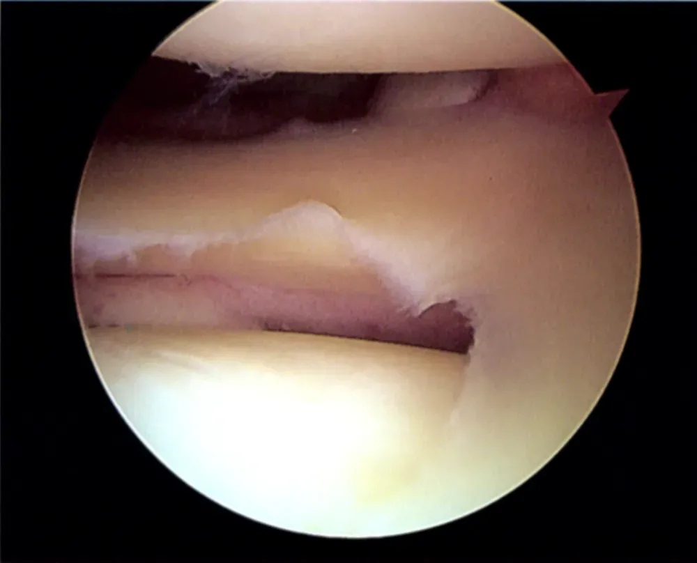
Knee surgeons used to operate under a blunt philosophy known informally as “if in doubt, take it out.” Any meniscus tear, no matter how minor, often led to a total meniscectomy – complete removal of the C-shaped cartilage that cushions the knee joint.
Biomechanical testing has since proven exactly how costly that approach was. The shock-absorbing capacity of a knee joint drops significantly once meniscal tissue is resected – the loss factor decreased by up to 68% after total meniscectomy compared to an intact knee.
That’s not a minor dip – that’s nearly two-thirds of the knee’s natural cushioning, gone. Total meniscectomy strips away the joint’s shock absorber entirely and raises the risk of osteoarthritis significantly more than a partial removal ever would.
Total meniscectomy is now a rare procedure, with surgeons instead fighting to preserve or repair as much tissue as possible. Some orthopedic specialists go further, arguing that decades of “quick fix” meniscus removals directly fueled today’s wave of early-onset knee arthritis in former athletes.
#9 – The Foreskin
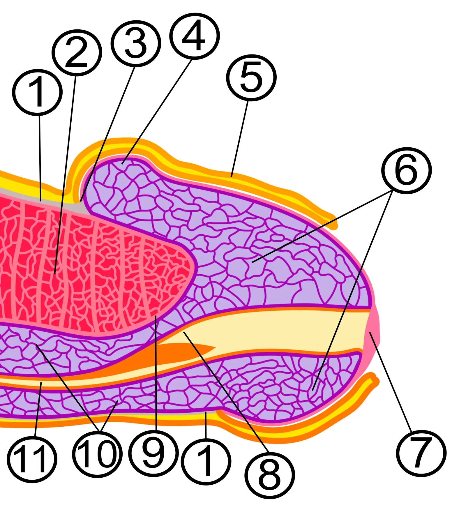
Few topics generate more heated debate than this one, and for good reason – circumcision has been practiced for religious, cultural, and medical reasons for thousands of years, often justified on the assumption that the foreskin serves no meaningful biological purpose.
Anatomically, it’s a mobile, double-layered fold of skin packed with a dense network of nerve endings and specialized touch receptors. It also produces natural lubrication and physically protects the glans, which becomes exposed to friction and keratinization once that covering is gone.
Most people don’t realize the foreskin contains some of the most nerve-dense tissue on the entire body, comparable in sensitivity to the fingertips or lips. Whether that translates to meaningful long-term differences in sensation is still fiercely debated, with studies producing mixed and often contradictory results.
What’s less debated is that it isn’t simply “extra skin” – it’s a functional structure with sensory and protective roles. That’s part of why many pediatric associations worldwide have shifted away from recommending routine, non-medical infant circumcision as a default practice.
Quick Compare
- Fingertips – dense receptor network built for fine touch discrimination
- Lips – highly innervated tissue used for sensory feedback
- Foreskin – anatomically comparable nerve density, per tissue studies
#10 – The Coccyx (Tailbone)
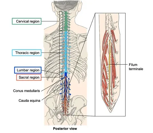
The tailbone has been mocked for generations as humanity’s most obvious leftover from our tree-swinging ancestors – a shrunken, fused remnant of a tail we supposedly no longer need. Coccygectomy, surgical removal of the tailbone, has occasionally been performed for chronic, treatment-resistant tailbone pain.
But surgeons who’ve operated in that area know the coccyx isn’t just dangling dead weight. It’s a critical anchor point – a dozen or more muscles, ligaments, and tendons attach directly to it, including pelvic floor muscles that support bladder and bowel control, and gluteal muscles involved in hip movement.
The tailbone also bears real mechanical load. It helps support your body weight while sitting, particularly when you lean back, acting almost like a third leg of a tripod alongside your two sitting bones.
Remove it, and the pelvic floor loses one of its structural anchors – which is exactly why coccygectomy is treated as a last-resort procedure with a notoriously long, difficult recovery, reserved only for the most severe cases.
#11 – The Palmaris Longus Muscle
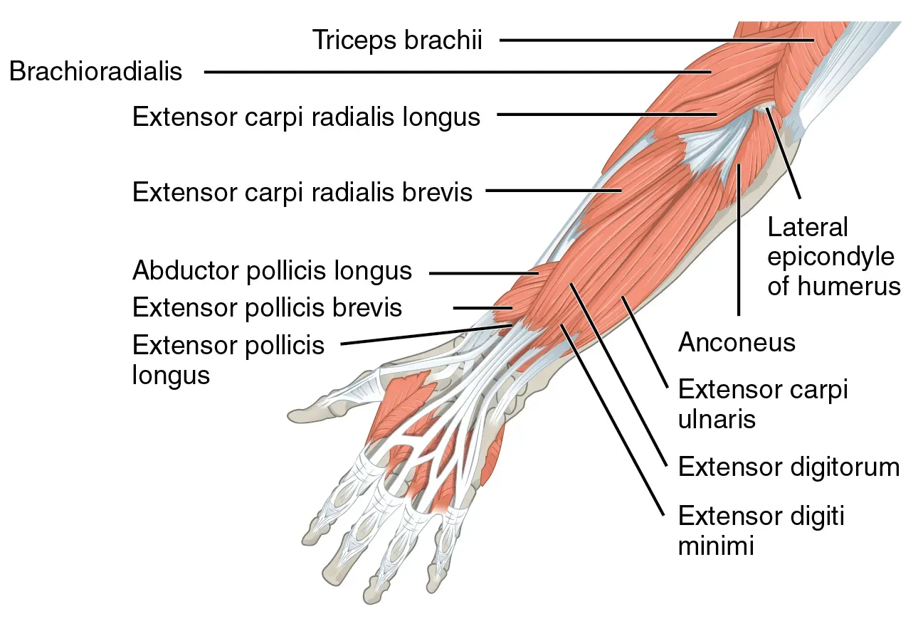
This one isn’t about doctors removing something out of caution – it’s about doctors actively harvesting it, because for decades the palmaris longus was considered so useless that surgeons treated it like a spare-parts bin.
Roughly 10 to 15% of people don’t even have this muscle in one or both forearms, and they notice zero difference in grip strength or wrist function. That absence alone convinced generations of anatomists it was a vestigial holdover doing nothing for modern humans.
Here’s the twist: its “uselessness” is exactly why it became medically valuable. Because the tendon can be removed with almost no functional loss to the donor site, surgeons routinely harvest it for reconstructive procedures – repairing torn ligaments elsewhere, rebuilding tendons in the hand, even reconstructing vocal cords and eyelids.
It’s a rare case where an organ’s uselessness became its defining medical utility, turning what was once dismissed as evolutionary leftover tissue into one of the most frequently borrowed materials in reconstructive surgery.
#12 – The Ovaries
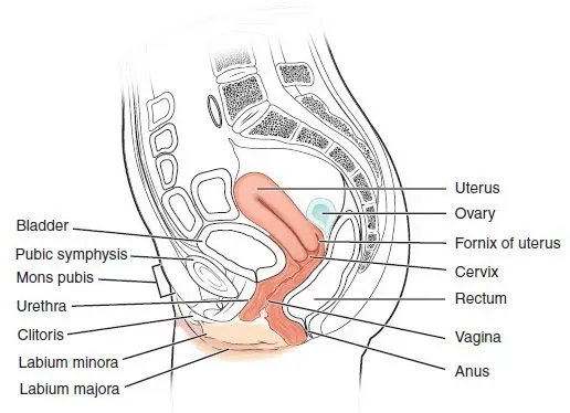
For much of the 20th century, when a woman underwent a hysterectomy, surgeons often removed her ovaries at the same time – even with no cancer or disease present. The logic was blunt: once childbearing was off the table, the ovaries had supposedly finished their job.
That reasoning ignored a crucial fact. The ovaries don’t just produce eggs – they’re a major hormone-producing gland, generating estrogen, progesterone, and a meaningful share of the body’s testosterone well beyond a woman’s reproductive years. Those hormones shape bone density, cardiovascular health, cognitive function, and mood for decades after fertility ends.
Removing healthy ovaries essentially triggers instant, surgical menopause – regardless of a woman’s actual age. Studies have linked premature ovarian removal to elevated long-term risks of heart disease, osteoporosis, and cognitive decline.
Medical guidelines have shifted significantly in response, now generally recommending ovarian conservation during hysterectomy for women without a genetic or cancer-related reason to remove them – a reversal that came only after decades of routine, preventive surgeries that likely did more harm than good.
At a Glance
- Ovaries produce estrogen, progesterone, and a share of the body’s testosterone
- Hormone output continues for decades beyond reproductive years
- Removal triggers immediate surgical menopause, at any age
- Linked to higher long-term risk of heart disease, osteoporosis, and cognitive decline
#13 – The Infrapatellar (Hoffa’s) Fat Pad
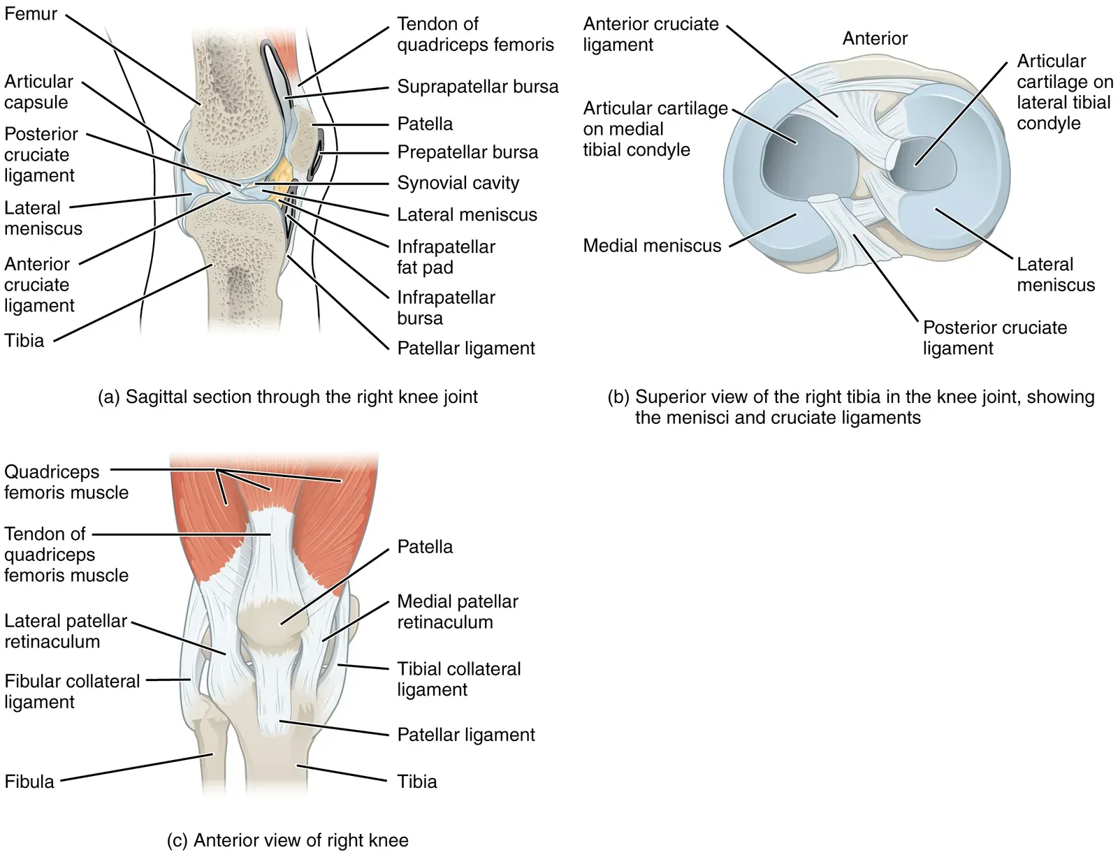
Rounding out this list is a piece of anatomy that surgeons have historically treated as nothing more than an annoying obstruction: the infrapatellar fat pad, sometimes called Hoffa’s fat pad, sitting just behind the kneecap.
For years, this soft tissue was routinely trimmed away or partially excised during arthroscopic knee procedures, simply because it got in the way of the surgeon’s tools and line of sight. It was written off as inert padding – filler tissue with no real biomechanical job.
Turns out, this fat pad is metabolically active and mechanically important. It cushions the knee joint, reduces friction between moving structures, and even secretes inflammatory and anti-inflammatory signaling molecules that influence joint health over time.
Excessive removal has since been linked to increased anterior knee pain and altered joint mechanics after surgery. Many arthroscopic surgeons have quietly shifted toward preserving as much of this tissue as possible – a small but telling admission that what looked like disposable padding was doing real structural and biochemical work all along.
The Bottom Line
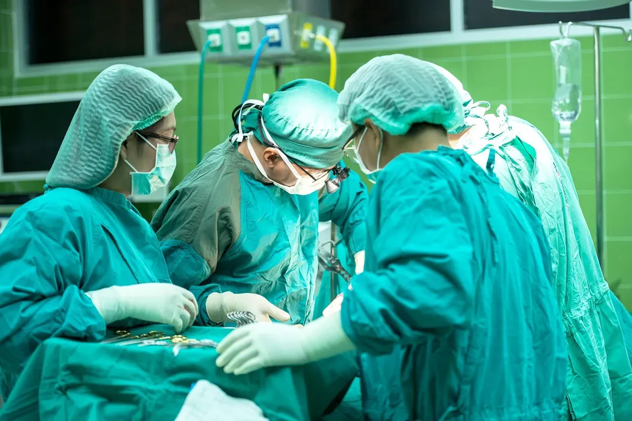
The pattern across all thirteen entries is impossible to ignore: medicine has a long, uncomfortable history of confusing “I don’t understand this organ’s function” with “this organ has no function.” From the appendix’s role as a bacterial safe house to the meniscus losing 68% of its shock-absorbing power after removal, the evidence keeps pointing the same direction – the body rarely builds anything truly pointless.
Surgery still saves lives when these structures are genuinely diseased, and none of this is an argument against necessary operations. But the routine, preventive removals of the past look increasingly reckless in hindsight, and they’re a humbling reminder that “we don’t know what it does” was never the same thing as “it doesn’t do anything.”
Which of these surprised you the most? Drop your take in the comments.