Think about it. Every fossil you’ve ever seen in a museum had to be painstakingly removed from rock, analyzed, and interpreted. For centuries, that meant hammers, chisels, and a whole lot of guesswork. Yet here’s the thing: the tools we use to study ancient life have transformed more in the past few decades than in the previous century combined. What was once impossible is now routine, and what seemed like science fiction is becoming everyday practice in paleontology labs around the world.
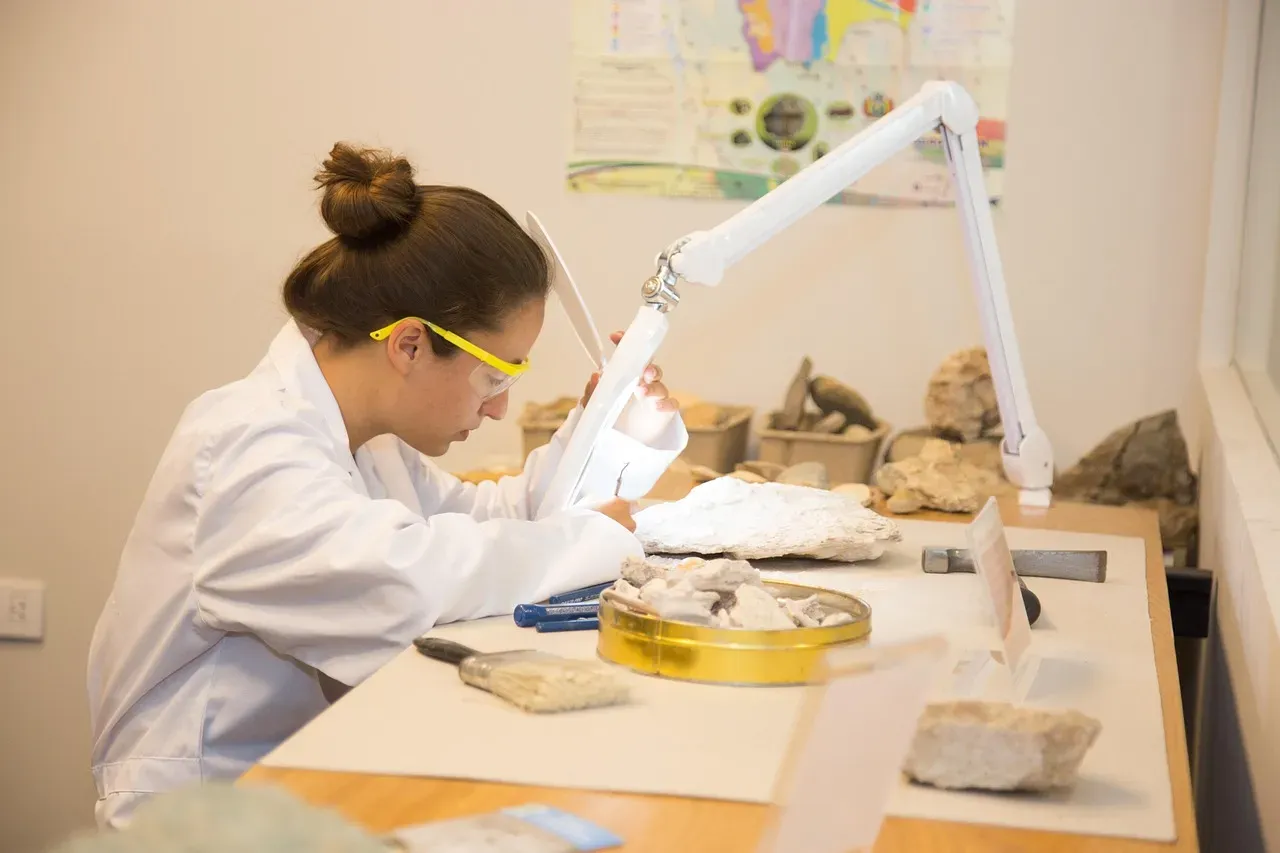
Let’s be real. You might imagine paleontology as dusty fieldwork and careful brushing, and honestly, that’s still a big part of it. Still, behind the scenes, researchers are deploying some of the most advanced technology available to unlock secrets that bones and rocks have kept hidden for millions of years. From seeing inside fossils without breaking them open to reading genetic codes from specimens older than you can fathom, the revolution is already here. So let’s dive in.
CT Scanning: Seeing Through Stone Without Breaking It
You can now create detailed 3D models of fossil neck vertebrae and skulls, analyzing bone structure in unprecedented detail to understand the biomechanics of massive dinosaurs. Here’s what makes this so remarkable: traditional methods would require physically cutting through these irreplaceable specimens, destroying them in the process. CT scanners reveal fossils’ internal structures, and advanced computer programs can analyze fossil data and reconstruct skeletons.
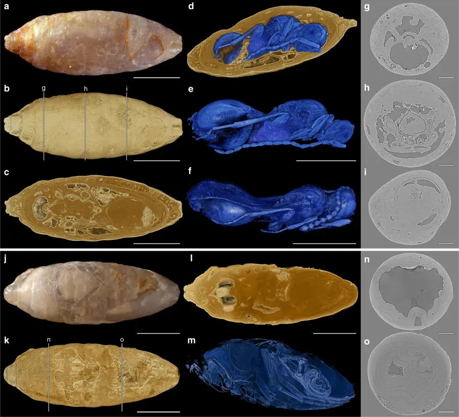
The technology has come a long way since its medical origins. CT has emerged as a widely used, nondestructive, and accurate tool for collecting fossil data, investigating the internal structure of fossils by acquiring thousands of serial images to produce accurate internal morphological 3D reconstructions. Imagine holding a fossil egg in your hand and being able to see the tiny bones of an embryo curled inside, all without cracking the shell. That’s not fantasy anymore. A 320-slice CT scanner allowed for a clear image of fossils despite the plaster cast surrounding them as protection from damage while in storage.
Deep Learning and Artificial Intelligence: Automating the Impossible
Honestly, the manual work involved in processing CT scans used to be mind-numbing. Picture this: you spend weeks, sometimes months, going through thousands of individual scan slices, painstakingly outlining where fossil ends and rock begins. Manual image segmentation is often the only way to digitally extract the regions of interest from CT slice stacks, and this process can take weeks to months to complete, being a considerable bottleneck in making data available for research.
Automated Deep Learning segmentation can obtain high-fidelity 3D models of fossils digitally extracted from the surrounding rock, training the model with less than roughly one to two percent of the total CT dataset, and this workflow has the capacity to revolutionize the use of Deep Learning to significantly reduce processing time. It sounds crazy, but neural networks can now learn what a fossil looks like versus the matrix around it. Deep learning can reduce processing time from days or weeks to minutes, although the 3D renderings from automated segmented slices are not as meticulous as manual results. The speed gain is absolutely game-changing for researchers who want to study specimens rather than spend their careers processing data.
Ancient DNA Extraction: Listening to Genetic Whispers
The petrous ear bone is most often used for DNA extraction, since its dense structure provides good conditions for DNA preservation. Yet even with ideal conditions, you’re working with fragments. Ancient DNA is more degraded compared to present-day genetic material due to degradation processes, and even under the best preservation conditions, there is an upper boundary of roughly half a million to one and a half million years for a sample to contain sufficient DNA for sequencing technologies.
Let’s be real about the challenges here. DNA is continuously being split up, and while the organism is alive these splits are repaired, but once an organism has died, the DNA will begin to deteriorate without repair, resulting in samples having strands of DNA measuring around a hundred base pairs in length. That’s incredibly short compared to modern DNA. The study of ancient genetic material is extremely difficult due to its poor quality and quantity, as well as possible contamination with modern DNA, and the most serious criticism in the paleogenetics field is the potential contamination of samples by contemporary DNA, since modern DNA consists of intact template molecules that will be copied with much higher efficiency during the PCR process than the fragmented and damaged ancient DNA templates. The precautions required are intense: dedicated clean rooms, full body protection, and obsessive protocols to prevent a single stray hair from ruining months of work.
Synchrotron Radiation: The Ultimate Microscope
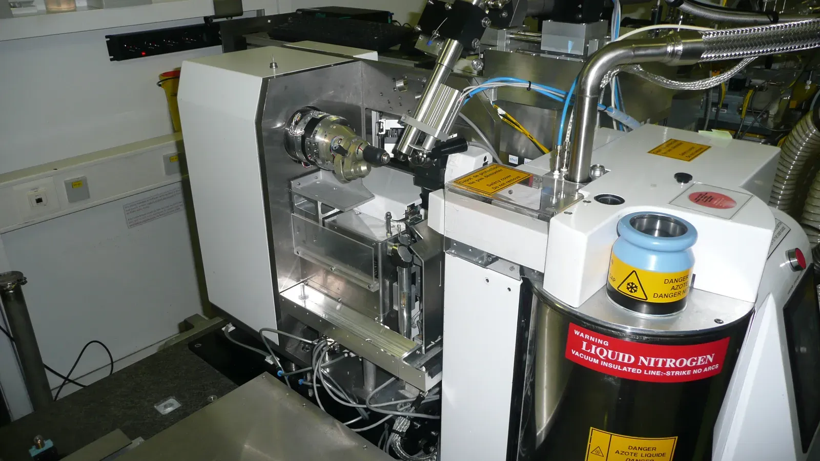
Think of synchrotron radiation as the Rolls Royce of imaging techniques. Synchrotron radiation is an electromagnetic radiation produced when electrons are forced to assume a curved trajectory resulting in an emission of a bright beam of high energy photons, carried out in large particle acceleration facilities called synchrotrons equipped with experimental stations at the end of each spot where the beam is emitted, and in an applied sense, synchrotron radiation allows the investigation of materials of different nature, from synthetic to biological, from macro to the nano-scale. These aren’t instruments you find in every university lab. They’re massive facilities, sometimes the size of several football fields.
Due to the high resolution and non-destructive properties, synchrotron techniques have provided invaluable information on rare and delicate fossils, enabling the detection and evaluation of the spatial distribution of trace elements like rare earth elements, and synchrotron X-ray fluorescence offers the opportunity of mapping the spatial distribution of elements with high sensitivity down to trace concentrations. Here’s what really gets me excited: The problem often in paleontology is that fossil bones and the surrounding rock have similar densities, thwarting the chance of identifying them via common nondestructive imaging methods, but phase-contrast microtomography is particularly sensitive to minute differences in the densities of a sample’s components. You can literally see things that are invisible to conventional methods.
Virtual Taphonomy: Predicting Fossilization Before It Happens

The ability to gain search images in both 2D and 3D for potential fossils gives paleobotanists a tool called virtual taphonomy to improve understanding of plant evolution and paleobiogeography. It’s hard to say for sure, but this might be one of the most underappreciated developments in recent years. Taphonomy is the study of how organisms decay and become fossils. Virtual taphonomy flips the script entirely.
Compared to CT, there is no beam-hardening effect that results in inaccurate portrayal of true X-ray absorption, and synchrotron X-ray tomographic microscopy has very high resolution, up to roughly a third of a micrometer, providing better resolution than high-resolution CT, and combined with recent developments in phase contrast methods, synchrotron facilities are a powerful tool for noninvasive volumetric investigation. Scientists can now take modern organisms, simulate decay processes digitally, and predict what kinds of features would survive fossilization. This helps them know what to look for in actual fossils and avoid misinterpreting what they find. Imagine being able to test your hypotheses about ancient soft tissue preservation without waiting millions of years. That’s the power here.
Non-Destructive DNA Recovery from Artifacts
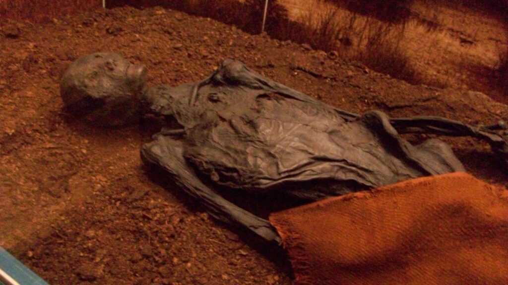
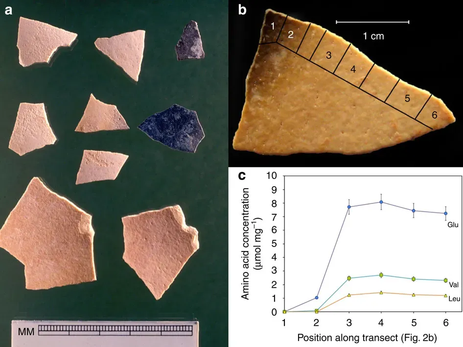
A non-destructive method for the gradual release of DNA trapped in ancient bone and tooth artefacts has been developed, and application of the method to an Upper Palaeolithic deer tooth pendant from Denisova Cave, Russia, resulted in the recovery of ancient human and deer mitochondrial genomes, allowing estimation of the pendant age at approximately nineteen thousand to twenty-five thousand years. Think about what this means: we’re not just studying bones anymore. We’re extracting genetic information from objects that ancient humans wore and touched.
The technique is genuinely groundbreaking. Stepwise extraction of DNA makes it possible to closely monitor the release of different DNA components during the extraction process, including endogenous DNA, environmental DNA from the surrounding sediment, ancient human DNA and present-day contamination, potentially allowing inferences about whether these components originate from traces of sediment that may still be adherent to the object, from its surface or its interior. The controlled, gradual approach means you can track exactly where the DNA is coming from at each stage. You’re not just getting genetic data; you’re getting spatial and contextual information too. The implications for understanding who made and used ancient artifacts are staggering.
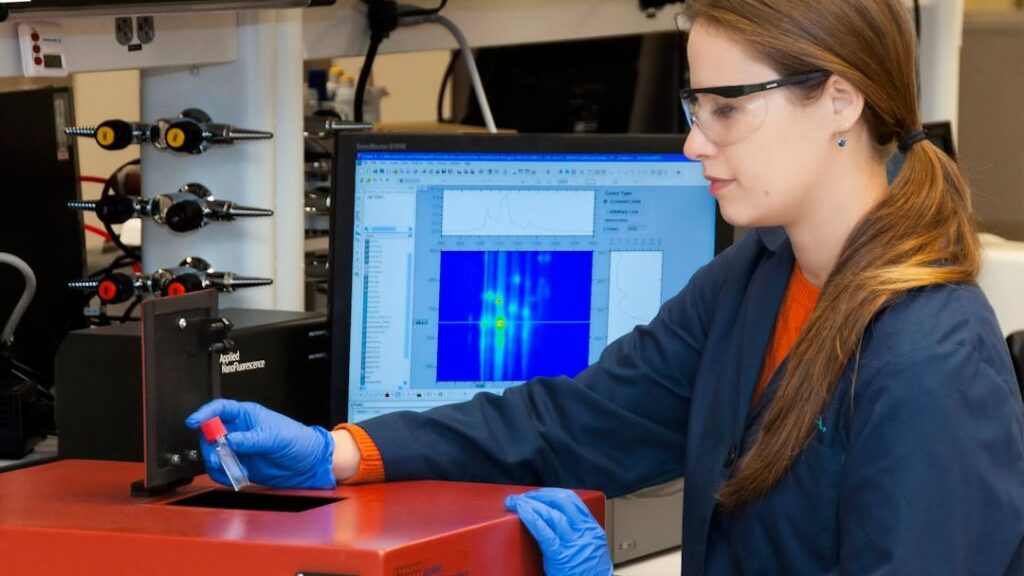
Molecular Paleontology: Reading Ancient Proteins and Biomolecules
Let’s be honest: DNA gets all the attention, but proteins and other biomolecules can tell us things DNA simply cannot. Researchers have uncovered thousands of preserved metabolic molecules inside fossilized bones millions of years old, offering a surprising new window into prehistoric life. These aren’t genes. They’re the actual chemical machinery that organisms used to live and function.
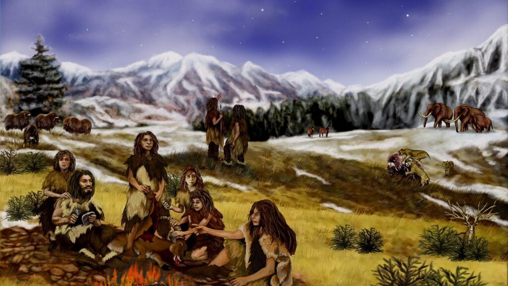
Scientists extracted and sequenced ancient RNA from thirty-nine thousand-year-old woolly mammoth tissues, a breakthrough because RNA degrades much faster than DNA and almost never fossilizes, marking one of the first successful recoveries of gene-expression material from deep time, and RNA reveals physiology, gene regulation, and cellular activity that DNA alone cannot show. RNA is incredibly fragile, far more so than DNA. Finding it in ancient specimens was thought to be basically impossible until very recently. The fact that researchers can now isolate and sequence it opens entirely new questions about ancient metabolism, development, and adaptation.
High-Precision Radiometric Dating from Fossils Themselves
Researchers have found that fossilized dinosaur eggshells contain a natural clock that can reveal when dinosaurs lived, and the technique delivers surprisingly precise ages and could revolutionize understanding. Dating fossils has always been tricky. Usually you date the rocks around them, not the fossils themselves. That introduces uncertainty and depends on having the right kind of surrounding material.
The breakthrough here is moving from relative dating to absolute dating using the fossils as primary sources. Traditional methods often relied on volcanic layers or other datable rock types nearby. When those aren’t present, you’re left with educated guesses based on which species are found together. Direct dating from fossilized material changes everything. You can establish precise timelines, test hypotheses about extinction events, and correlate finds from different continents with confidence you simply couldn’t have before. The development of radiometric dating allowed absolute dates to be assigned to the geologic timescale, and the theory of plate tectonics helped make sense of the geographical distribution of ancient life. Together, these advances have revolutionized how we understand the history of life on Earth.
Conclusion
Here’s the thing. These eight breakthroughs aren’t just making paleontology faster or easier. They’re fundamentally changing what questions we can even ask about ancient life. We’ve moved from describing what extinct organisms looked like to understanding how they moved, what they ate, how they grew, who their descendants are, and even what molecules coursed through their bodies millions of years ago.
What would you have guessed was possible fifty years ago? Probably not extracting readable DNA from bones tens of thousands of years old. Definitely not seeing inside a fossil egg without breaking it, or teaching a computer to recognize fossils in CT scans. The boundaries keep shifting, and honestly, that’s what makes this field so exhilarating right now. Every technical advance reveals layers of information that were always there, just waiting for us to develop eyes capable of seeing them.